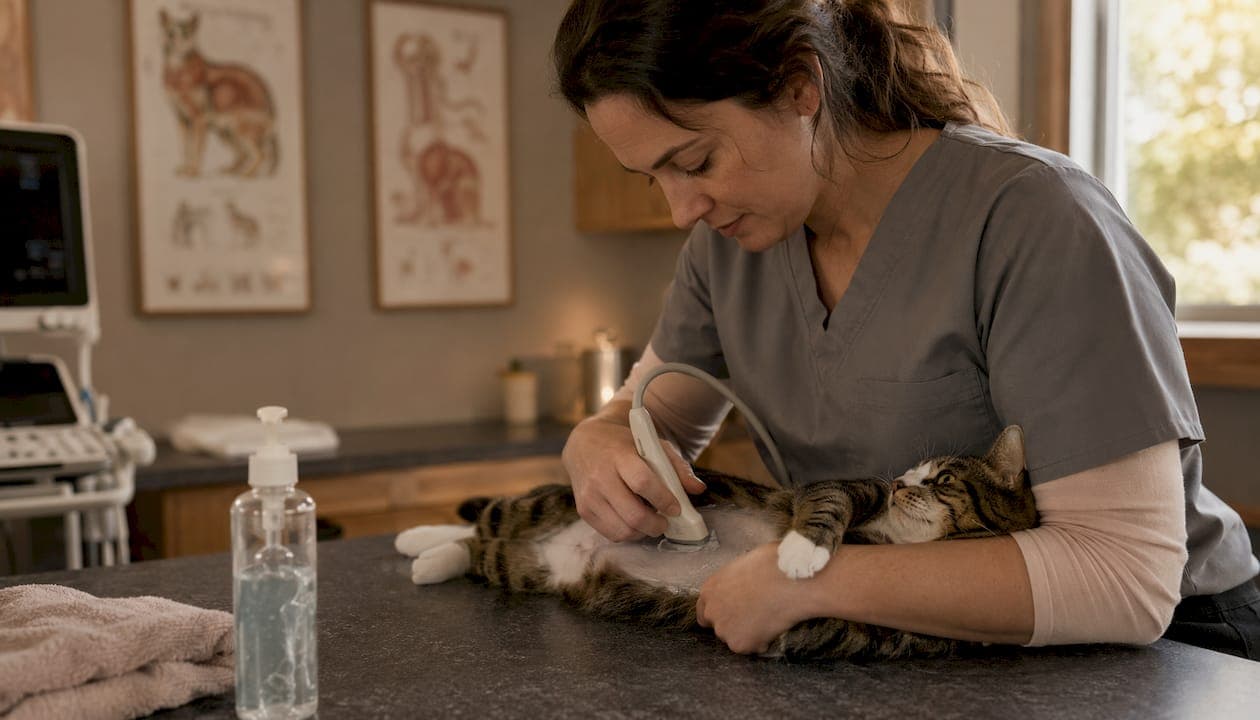
Transabdominal ultrasound can confirm a cat pregnancy as early as day 11 (embryonic vesicle) and detect fetal heartbeats by approximately day 16–18, with the most reliable diagnostic window falling between days 25 and 35. Scans performed before day 21 carry a real risk of false negatives. Three things ultrasound does well: it confirms pregnancy, assesses fetal viability, and guides recheck scheduling. Three things to know upfront:
- Confirmation: Embryonic vesicles appear around day 11–16; embryos are visible by day 15–17.
- Viability: Fetal heartbeats, assessed with M-mode or Doppler, are typically visible by day 16–18 and are the primary viability marker.
- Limitations: Ultrasound is a poor tool for counting fetuses. The Merck Veterinary Manual recommends using radiographs after skeletal ossification for accurate litter counts.
Table of Contents
- What does ultrasound show at each stage of cat pregnancy?
- How is a cat pregnancy ultrasound actually performed?
- How do vets estimate gestational age from measurements?
- How accurate is ultrasound, and where does it fall short?
- What other methods can confirm or monitor a cat pregnancy?
- Why does the operator's skill matter so much?
- What does a cat pregnancy ultrasound cost in the U.S.?
- Key Takeaways
- Why ultrasound is worth doing right, not just doing fast
- Pregnancy ultrasound for your cat at Cityanimalvet
- Useful sources
What does ultrasound show at each stage of cat pregnancy?
The sonographic picture changes quickly across gestation. Knowing what to expect at each stage tells you whether a scan is worth doing now or whether waiting a week will give you far more information.
| Gestational stage (rough) | Expected sonographic finding | Clinical action |
|---|---|---|
| Early embryonic period | Embryonic vesicle (anechoic fluid sac) | Confirm pregnancy; recheck if inconclusive |
| Mid embryonic period | Embryo visible within vesicle | Confirm embryo presence |
| Early fetal period | Fetal heartbeat detectable (M-mode/Doppler) | Assess viability |
| Optimal window | Head, stomach, limb buds visible; best for dating | Biometric measurements |
| Later fetal period | Skeletal ossification begins | Switch to radiographs for fetal counting |
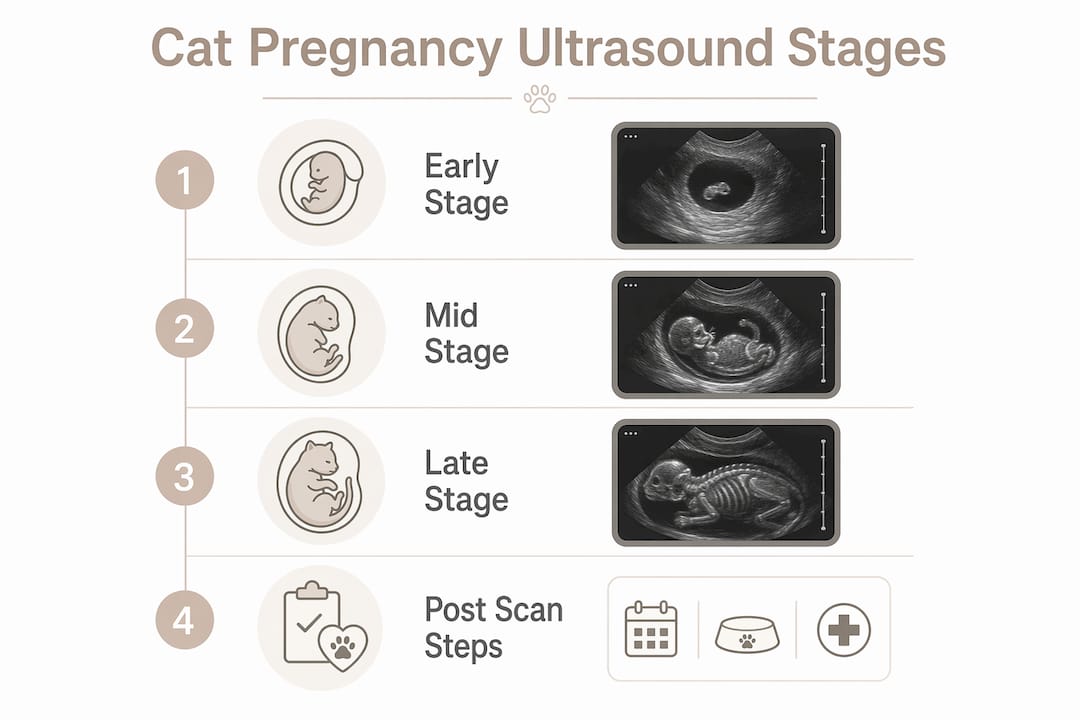
A 12.5 MHz probe study confirmed vesicles from day 11, embryos from day 15–17, and heartbeats from day 16–18. Higher-frequency probes improve resolution for early structures but sacrifice depth penetration, so probe selection should match the cat's body size and the gestational stage.
Key practical points about the timeline:
- Scanning before day 21 risks a false negative.
- The days 25–35 window is optimal for both confirming pregnancy and obtaining reliable biometric measurements.
- Heartbeat detection is the single most important viability indicator at any stage.
- Ossification around day 42 is the trigger to shift from ultrasound to radiographs for counting.
How is a cat pregnancy ultrasound actually performed?
The exam is straightforward for a calm, cooperative cat, and most queens tolerate it without sedation. Here is what happens from check-in to final documentation.
Preparation and positioning: The cat arrives with a recent history (mating date if known, any medications, prior reproductive issues). Hair clipping over the caudal abdomen is done only when the coat is thick enough to degrade image quality. Most cats are placed in dorsal recumbency, though lateral recumbency works equally well. The University of Florida imaging resource notes that both positions are effective for abdominal scanning, and the choice often comes down to what keeps the patient calmest.
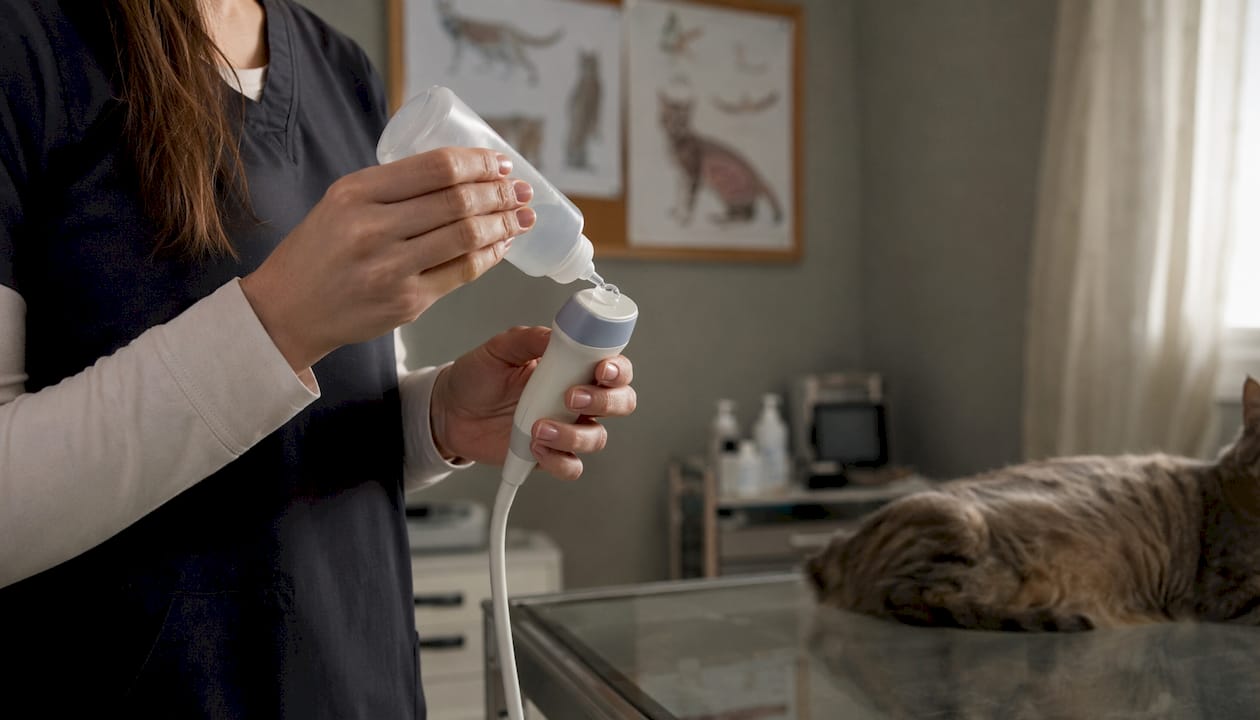
Probe selection and technique: Probes in the 7.5–12.5 MHz range are standard for feline reproductive imaging. Higher frequencies (10–12.5 MHz) give better resolution for early embryonic structures; lower frequencies (5–7.5 MHz) are used for larger or obese cats where depth penetration matters more. The sonographer sweeps both uterine horns systematically, angling and rotating the transducer to visualize each gestational sac in two planes.
What gets documented: Per ACVR/ECVDI consensus guidance, a thorough exam records still images of both uterine horns, representative gestational sacs, individual fetuses, and an M-mode or Doppler clip confirming fetal heart rate. Fetal heart rate in kittens runs approximately two to three times the dam's rate, making it a reliable viability marker distinct from maternal cardiac activity.
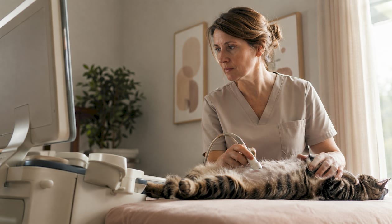
Owner logistics: Most feline pregnancy scans take 20–40 minutes. No fasting is required. Owners should bring any records of the mating date and note whether the cat is on any medications. Results are typically delivered as still images, short video clips, and a written measurement report.
Pro Tip: When performing M-mode for fetal heart rate, freeze the image and scroll back rather than trying to capture a moving cursor in real time. This gives a cleaner waveform and a more defensible measurement for the medical record.
How do vets estimate gestational age from measurements?
Fetal biometry turns a pregnancy scan into a dating tool, which matters for predicting parturition and planning any intervention. Three measurements do most of the work.
Common measurements:
- Embryonic vesicle diameter (EVD): Useful in the first two weeks; correlates with gestational age before the embryo is clearly visible.
- Crown-rump length (CRL): Measured from the top of the head to the base of the tail; most reliable in the first half of gestation.
- Biparietal diameter (BPD): The width of the fetal skull measured at its widest point; the most widely validated measurement for estimating days to parturition in the second half of gestation.
The Animal Ultrasound Association publishes the Beccaglia formula for BPD-based dating:
Experienced operators using this formula can expect accuracy within a few days. That precision depends on technique: the BPD is measured in a true transverse plane through the thalamus.
| Measurement | Gestational stage | Typical range | Formula |
|---|---|---|---|
| Embryonic vesicle diameter | Day 11–20 | 2–10 mm (early) | Correlates with early days; no single published formula |
| Crown-rump length | Day 20–35 | 5–35 mm | Used for early dating; less reliable late |
| Biparietal diameter | Day 30–42 | 10–30 mm | Days to parturition = (BPD mm − 23.39) / 0.47 |
The Animal Ultrasound Association recommends measuring at least three fetuses, ideally on different kittens, and averaging the results. A single measurement from one fetus introduces too much biological variability to be reliable, especially in larger litters where growth rates can differ.
How accurate is ultrasound, and where does it fall short?
Ultrasound is excellent at two things: confirming that a pregnancy exists and assessing whether fetuses are alive. It is genuinely poor at one thing: counting how many fetuses there are.
Where false negatives happen:
- Scanning before day 21, when vesicles may be too small to resolve clearly.
- Using a low-frequency probe (5 MHz or below) in early gestation.
- Operator inexperience with feline reproductive anatomy.
- Resorption of one or more embryos, which can make a litter appear smaller or absent.
The Merck Veterinary Manual is direct: scans before day 21 may give false negatives, and the optimal window is days 25–35. If a scan at day 18 is negative but the clinical picture suggests pregnancy, a recheck at day 25 is the right call, not a conclusion that the cat is not pregnant.
Fetal counting: The University of Florida imaging resource states plainly that sonography provides a poor method for counting fetuses. Gestational sacs overlap, fetuses move, and the uterine horns curve in ways that make it easy to count the same fetus twice or miss one entirely. Radiographs after ossification around day 42 are the standard for determining actual litter size when that number matters clinically.
Viability assessment is where ultrasound earns its keep. A fetal heart rate visible on M-mode is the clearest sign of a live fetus. No heartbeat on a well-performed scan after day 18 is a strong indicator of fetal death, though a recheck 24–48 hours later is prudent before drawing a definitive conclusion.
Pro Tip: Document fetal heart rate with a saved M-mode clip at every pregnancy scan, not just when viability is in question. That baseline becomes invaluable if the owner returns with concerns about fetal loss a week later.
What other methods can confirm or monitor a cat pregnancy?
Ultrasound is the most versatile tool, but it works best alongside other diagnostics rather than in isolation.
Abdominal palpation: An experienced clinician may detect uterine enlargement or discrete gestational swellings around day 21–25, but reliability drops sharply in obese cats, anxious cats, or large litters where individual sacs are hard to distinguish. Palpation cannot confirm viability and should never be the sole diagnostic method.
Relaxin assay: A blood test measuring relaxin, a hormone produced by the feline placenta, becomes positive after approximately day 25–30. It confirms pregnancy but gives no information about viability, litter size, or gestational age. It is a useful adjunct when ultrasound is unavailable or inconclusive.
Radiographs: The go-to tool after day 42, when fetal skeletons are sufficiently ossified to count individual skulls and spines reliably. Radiographs are the standard for pre-whelping litter counts and for evaluating fetal positioning when dystocia is a concern.
| Method | Earliest reliable timing | Best use case | Limitations |
|---|---|---|---|
| Ultrasound | Day 21–25 (optimal day 25–35) | Confirmation, viability, dating | Poor for counting; operator-dependent |
| Abdominal palpation | Day 21–25 | Initial screening | Unreliable; no viability data |
| Relaxin assay | Day 25–30 | Confirm pregnancy when ultrasound unavailable | No viability or dating information |
| Radiographs | Day 42+ | Fetal counting, dystocia planning | Radiation; no viability data |
Practical scenarios:
- Single unintended mating: Ultrasound at day 25–28 confirms pregnancy and viability; relaxin test as backup if scan is inconclusive.
- Suspected fetal loss: Repeat ultrasound 24–48 hours after initial concern; compare fetal heart rates and sac number.
- Planning for C-section: Radiograph at day 45–50 for accurate count; ultrasound closer to term to assess fetal viability and position.
Why does the operator's skill matter so much?
Ultrasound is not a passive technology. The image on the screen is only as good as the person holding the probe, and feline reproductive imaging has specific technical demands that general practice experience does not always cover.
Operator-dependent variables include:
- Probe angle and pressure, which affect whether a small embryonic vesicle is visible or missed entirely.
- Measurement reproducibility: BPD measurements taken in a slightly oblique plane can shift the dating estimate by several days.
- Distinguishing resorption artifacts from live but small fetuses.
- Recognizing concurrent uterine pathology (pyometra, uterine cysts) that can mimic or coexist with pregnancy.
Referral criteria: Request a board-certified imaging specialist or a comprehensive abdominal scan when:
- Fetal viability is unclear after two scans.
- Measurements are inconsistent across fetuses or between examiners.
- Clinical history or physical exam suggests concurrent abdominal pathology.
- The pregnancy is high-risk (prior dystocia, uterine abnormalities, planned C-section).
- The initial scan was performed with a low-frequency probe or by an operator with limited reproductive imaging experience.
The ACVR/ECVDI consensus statement recommends a standardized, comprehensive abdominal ultrasound rather than a focused pregnancy-only scan when clinical history warrants it. A pregnancy check that misses a concurrent pyometra or abdominal mass is not a complete exam. Knowing when to look beyond the uterus is part of what separates a thorough scan from a quick confirmation.
What does a cat pregnancy ultrasound cost in the U.S.?
Cost varies considerably depending on clinic type, geographic region, and the scope of the exam. A focused pregnancy confirmation scan at a general practice typically runs less than a comprehensive abdominal ultrasound or a specialist-performed study. According to Catster's cost guide, prices vary widely by region and clinic, and owners should expect higher costs when a board-certified radiologist performs or reviews the scan.
Factors that raise the cost:
- Specialist or referral center vs. general practice.
- Comprehensive abdominal scan vs. focused reproductive exam.
- Geographic location (urban markets like New York City typically run higher).
- Whether sedation is needed for an uncooperative cat.
- House call or mobile ultrasound services.
Appointment checklist for owners:
- Note the mating date if known, even an approximate one.
- Bring a list of current medications.
- No fasting required for most ultrasound exams.
- Arrange transport that minimizes stress; a covered carrier with familiar bedding helps.
- Ask in advance whether the clinic offers Fear Free handling or house call options if your cat is particularly anxious.
Results are typically provided as still images, short video clips of fetal heartbeats, and a written report with measurements. Most clinics schedule a follow-up scan or recommend one at a specific gestational milestone, particularly if the initial scan was early or inconclusive.
Key Takeaways
Ultrasound is the most reliable tool for confirming feline pregnancy and assessing fetal viability, with the optimal diagnostic window falling between days 25 and 35 of gestation.
| Point | Details |
|---|---|
| Optimal scan timing | Days 25–35 give the most reliable confirmation and biometric data; before day 21 risks false negatives. |
| Viability assessment | Fetal heartbeats visible by day 16–18 via M-mode or Doppler are the primary viability marker. |
| Fetal counting limitation | Ultrasound is unreliable for counting fetuses; use radiographs after ossification around day 42. |
| Biometry accuracy | The Beccaglia BPD formula yields ±2 days accuracy when three or more fetuses are measured and averaged. |
| Cityanimalvet | Cityanimalvet in Brooklyn offers diagnostic ultrasound with Fear Free handling and house call options for anxious cats. |
Why ultrasound is worth doing right, not just doing fast
The conventional wisdom in busy general practice is that a quick pregnancy confirmation scan is enough: find a heartbeat, confirm the pregnancy, send the owner home. That approach works most of the time. But it misses the cases where it matters most.
A cat that presents for a pregnancy check may also have a concurrent uterine abnormality, an early pyometra developing alongside a pregnancy, or a litter where one or two fetuses have already resorbed. A focused scan that finds a heartbeat and stops there will not catch any of that. The ACVR/ECVDI consensus exists precisely because abbreviated scans, while convenient, carry real diagnostic risk when clinical history suggests something more complex.
The other underappreciated factor is handling. A cat that is stressed, vocalizing, and trying to escape the table produces worse images than a calm cat in a quiet room. Fear Free handling is not just a marketing phrase. It directly affects image quality, measurement reproducibility, and the owner's willingness to come back for the follow-up scan that often matters more than the first one.
Ultrasound is a tool. Its value is determined almost entirely by the skill of the person using it and the conditions under which it is performed. Owners deserve to know that, and clinicians deserve the time and environment to do it properly.
Pregnancy ultrasound for your cat at Cityanimalvet

Cityanimalvet in Brooklyn offers diagnostic ultrasound for pregnant cats with the same Fear Free approach that defines every visit at the practice. For cats that tense up the moment they enter a clinic, that difference shows up directly in image quality and in how much useful information comes out of the appointment. The team documents fetal heartbeats, takes biometric measurements, and provides a written report with images you can keep for your records.
For cats that find travel stressful, house call ultrasound is available, bringing the exam to your home and eliminating the carrier-and-car stress that can make a cat nearly impossible to scan. Whether you need an early confirmation scan, a follow-up viability check, or a comprehensive abdominal exam to rule out concurrent pathology, the clinic can tailor the appointment to where your cat is in her pregnancy.
Book a pregnancy ultrasound appointment at Cityanimalvet and ask about house call availability when you schedule.
Useful sources
For owners and clinicians who want to go deeper, these are the primary references behind this article:
- Merck Veterinary Manual: Pregnancy Determination in Bitches and Queens — The standard clinical reference for timing, method comparison, and false-negative risk. Best for both owners and clinicians.
- ACVR/ECVDI Consensus Statement on Abdominal Ultrasound — Specialist-level guidance on standardized documentation, required images, and when to perform a comprehensive rather than focused scan. Best for clinicians and sonographers.
- Animal Ultrasound Association: Calculating Gestational Age in Cats — Practical guide to the Beccaglia BPD formula, measurement technique, and expected accuracy. Best for clinicians performing biometry.
- Early Pregnancy Diagnosis with a 12.5 MHz Probe (J Feline Med Surg, 2014) — Peer-reviewed study establishing day ranges for vesicle, embryo, and heartbeat detection by probe frequency. Best for clinicians.
- University of Florida Veterinary Imaging Resource: Ultrasound of the Small Animal Abdomen — Comprehensive imaging reference covering technique, fetal counting limitations, and systematic scanning approach. Best for clinicians and students.
- Catster: What's the Price of an Ultrasound for a Cat? — Consumer-facing cost guide with regional variability context. Best for owners budgeting for the procedure.
This article is general veterinary information, not a substitute for professional advice. Confirm timing and approach with your veterinarian based on your cat's specific history and clinical presentation.