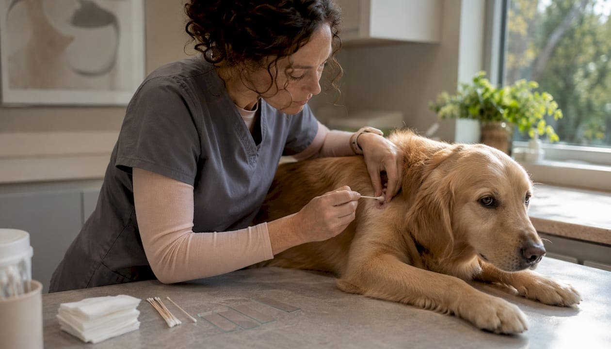
Skin cytology in dogs is one of the fastest, most cost-effective diagnostic moves available in a general practice setting. Press a slide to a pustule, stain with Diff-Quik, scan at 40x, then confirm at 100x oil immersion, and you can distinguish septic pyoderma from sterile pustular disease, bacterial overgrowth from Malassezia dermatitis, and inflammatory from neoplastic processes, all before the client leaves the room. The immediate clinical action is straightforward: collect the appropriate sample (impression smear, tape prep, or swab), stain with Diff-Quik or Gram stain when bacterial morphology matters, and act on what you see. Neutrophils packed with intracellular cocci mean pyoderma; abundant peanut-shaped yeast with minimal inflammation means Malassezia overgrowth. The World Association for Veterinary Dermatology recommends cytology at every otitis consult and recheck, and Cityanimalvet's Fear Free and home-visit services make this standard achievable even for anxious patients.
Key clinical actions at a glance:
- Collect sample by the method that matches the lesion: impression smear for erosions and pustules, tape prep for dry/scaly skin, swab for ear canals.
- Stain with Diff-Quik for rapid results; add Gram stain when bacterial morphology (cocci vs. rods) needs clarification.
- Scan at low power (10x) for distribution and quality, then 40x for semiquantitative cell counts, then 100x oil immersion for organism identification and intracellular detail.
- Treat or escalate based on the combination of cell type, organism density, and clinical signs, never on organisms alone.
Table of Contents
- When should you perform skin or ear cytology?
- How to collect diagnostic samples for maximum yield
- Staining and slide preparation: Diff-Quik, Gram, and heat-fix rules
- Microscopy workflow, magnifications, and a reporting template
- How to interpret cellular findings: neutrophils, eosinophils, mast cells, and more
- Recognizing bacteria, Malassezia, fungi, and ectoparasites on cytology
- Common cytologic patterns and what they mean for treatment
- Pitfalls and when to escalate beyond cytology
- Practical in-clinic workflow: cost, turnaround, and talking to owners
- Minimizing stress during sampling: Fear Free and home-visit adaptations
- Key Takeaways
- Why routine cytology changed how I practice dermatology
- Cityanimalvet's dermatology services: what to expect at your appointment
- Useful sources and further reading
When should you perform skin or ear cytology?
Cytology belongs on the diagnostic checklist for nearly every dermatology case at first consult, and again at every recheck until the slide is clean. The highest-priority indications are otitis externa (both ears, every patient, every visit), pustular or vesicular lesions, erosions and ulcers, pruritic erythematous skin, crusts or epidermal collarettes, interdigital lesions, and nodules or draining tracts.
For most acute presentations, cytology alone is sufficient to guide initial therapy. Culture becomes necessary when rods appear on cytology, when infection recurs after appropriate treatment, when multidrug resistance is suspected, or when otitis media is possible. Biopsy is the right next step for nodules that do not resolve, chronic unresolved dermatitis, or any lesion where neoplasia is on the differential.
A practical triage rule: perform cytology before culture whenever possible. Cytology is the most reliable rapid test for routine management of canine otitis and skin lesions, and it gives you actionable data in minutes rather than days. Culture without cytology often leads to treating a sensitivity report that does not match what is actually happening on the slide.
How to collect diagnostic samples for maximum yield
Technique determines whether a slide is diagnostic or a waste of stain. Match the collection method to the lesion type.
- Direct impression smear. Press a clean glass slide firmly against an erosion, ulcer, or moist lesion. Apply moderate pressure and lift straight off. This works well for exudative surfaces and gives you both cells and organisms in their natural ratio.
- Tape preparation. Press a strip of clear acetate tape against dry, scaly, or greasy skin, then mount it adhesive-side down on a drop of Diff-Quik blue stain on a slide. Adhesive slides capture more keratinocytes and surface debris, making tape prep the method of choice for seborrheic lesions and interdigital skin. Plain glass slides, by contrast, recover more extracellular bacteria when bacterial detection is the primary goal.
- Swab technique for ear canals. Insert a cotton-tipped swab into the horizontal ear canal, rotate gently, then roll (do not smear) the swab across a glass slide in a single direction. Rolling preserves cell morphology; smearing destroys it. Use a separate swab for each ear, because the two canals can harbor entirely different microbial populations.
- Pustule sampling. Open an intact pustule with a sterile needle tip and express the contents directly onto a slide. This is the single most diagnostic sample for pyoderma because it captures cells and organisms before secondary contamination occurs.
- Crust sampling. Peel the crust gently and press the slide against the moist base, not the crust surface. The base contains the active inflammatory cells; the crust itself is often acellular debris.
- Fine needle aspiration for nodules. Insert a 22–25 gauge needle into the nodule, redirect several times without withdrawing, then expel contents onto a slide with a syringe. Prepare multiple slides from a single lesion if the first attempt is non-diagnostic.
Site-specific notes: For interdigital lesions, tape prep or an adhesive slide works better than impression alone because the skin surface is often dry between flares. For lip fold and skin fold lesions, a direct impression or swab captures the moist exudate effectively.
Avoid contamination from clipped hair or topical products. Label slides immediately with patient ID, site, and date. Preparing two or three slides per lesion using different techniques costs almost nothing and dramatically reduces the chance of a non-diagnostic result.
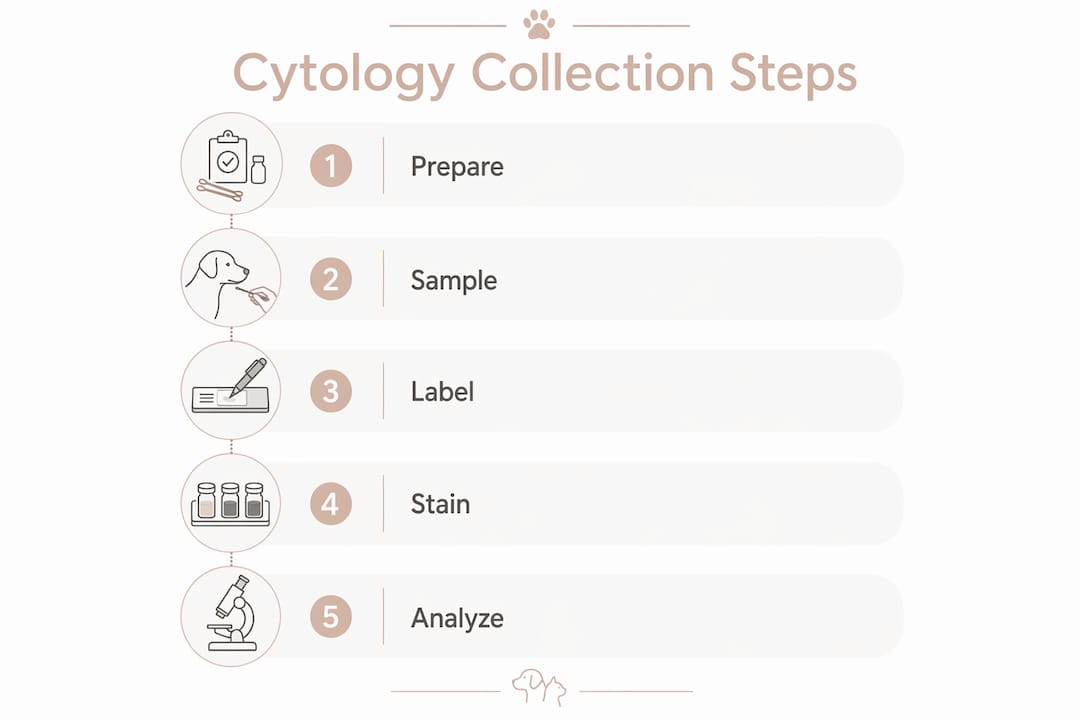
Pro Tip: Assemble a dedicated cytology kit tray: glass slides, adhesive tape strips, Diff-Quik reagents, Gram stain reagents, sterile cotton swabs, 22-gauge needles, and pre-printed labels. Keeping everything in one tray cuts setup time to under a minute and keeps technique consistent across technicians.
Staining and slide preparation: Diff-Quik, Gram, and heat-fix rules
Diff-Quik is the workhorse stain for in-clinic cytology. It stains nuclei blue-purple and cytoplasm pink-blue, differentiates leukocyte types clearly, and highlights Malassezia yeast and bacterial morphology well enough for most clinical decisions. The full three-step protocol (fixative, eosinophilic solution, basophilic solution) takes under two minutes.
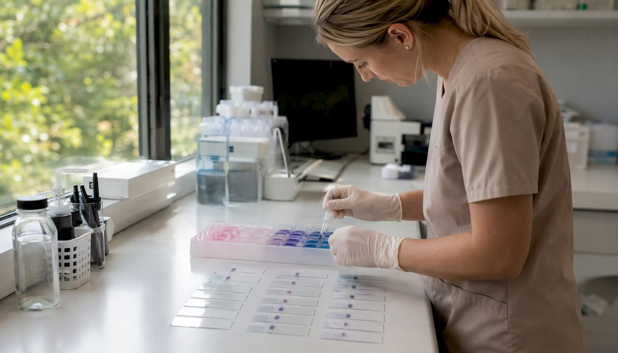
Air-dry before staining. Direct smears must be fully air-dried before Diff-Quik; residual moisture causes cells to lyse and organisms to wash off. Tape preparations go straight into the stain without drying because the tape itself holds cells in place.
Heat-fix rules. Heat-fix dry or greasy direct smears before staining to improve cell adhesion. Do not heat-fix moist samples or tape preparations. Heat on a moist slide destroys cellular morphology and reduces diagnostic value. The one-line rule: if the slide feels tacky or the sample came from a wet surface, skip the heat step entirely.
Gram stain adds value when you need to confirm bacterial morphology, particularly when rods are suspected or when the Diff-Quik result is ambiguous. Gram-positive cocci (Staphylococcus) stain purple; Gram-negative rods (Pseudomonas, Proteus) stain pink. This distinction matters for empirical antibiotic selection before culture results return.
Wright-Giemsa is an alternative to Diff-Quik that provides slightly better nuclear detail and is preferred by some reference laboratories for mast cell and lymphocyte differentiation. It is less practical for in-house use because of longer staining times.
Pro Tip: Overstaining is the most common in-clinic artifact. If slides consistently look too dark, reduce dip time in the basophilic solution by 30 seconds. Rinse with distilled water, not tap water, to avoid mineral deposits that mimic organisms at low power.
Microscopy workflow, magnifications, and a reporting template
Start every slide at low power (10x objective) to assess overall cellularity, staining quality, and distribution. Look for areas where cells are in a monolayer rather than clumped, because clumped areas distort morphology and inflate organism counts. Move to 40x (high dry) for semiquantitative leukocyte and organism counts. Finish at 100x oil immersion for organism identification, intracellular detail, and nuclear morphology.
Scan using a "battleship" pattern: move the slide in parallel horizontal rows, advancing one field width at a time, to cover the whole smear without revisiting areas. Scanning with this movement avoids over-reliance on the most cellular but potentially unrepresentative zones where organisms may be obscured by cell debris.
Semi-quantitative thresholds provide a starting framework, but they require clinical correlation. Counts above 5 yeasts or 25 bacteria per 40x field often suggest infection, but clinical signs must also be present to confirm a clinically significant diagnosis. Overlap exists between healthy and diseased dogs, so decisions should be based on organism density combined with clinical findings.
Cytology reporting fields
| Field | Example entry |
|---|---|
| Specimen type and site | Ear canal swab, right horizontal canal |
| Collection technique | Cotton swab, rolled on glass slide |
| Slide quality | Adequate cellularity, good staining, no significant artifact |
| Cell types (semiquantified) | Moderate degenerate neutrophils; rare macrophages |
| Organisms (type, location) | Numerous cocci, intracellular within neutrophils |
| Clinical interpretation | Consistent with septic otitis externa |
| Recommended next steps | Systemic antibiotics; recheck cytology in 3 weeks |
Document slide quality explicitly. A non-diagnostic slide due to poor sampling technique is a different clinical situation than a genuinely acellular lesion, and the distinction matters for follow-up planning.
Key documentation points:
- Record specimen type, site, and collection method every time.
- Note staining quality and whether artifacts limit interpretation.
- Semiquantify cells and organisms (rare, few, moderate, numerous) rather than using vague descriptors.
- State the clinical interpretation and the recommended next step clearly.
How to interpret cellular findings: neutrophils, eosinophils, mast cells, and more
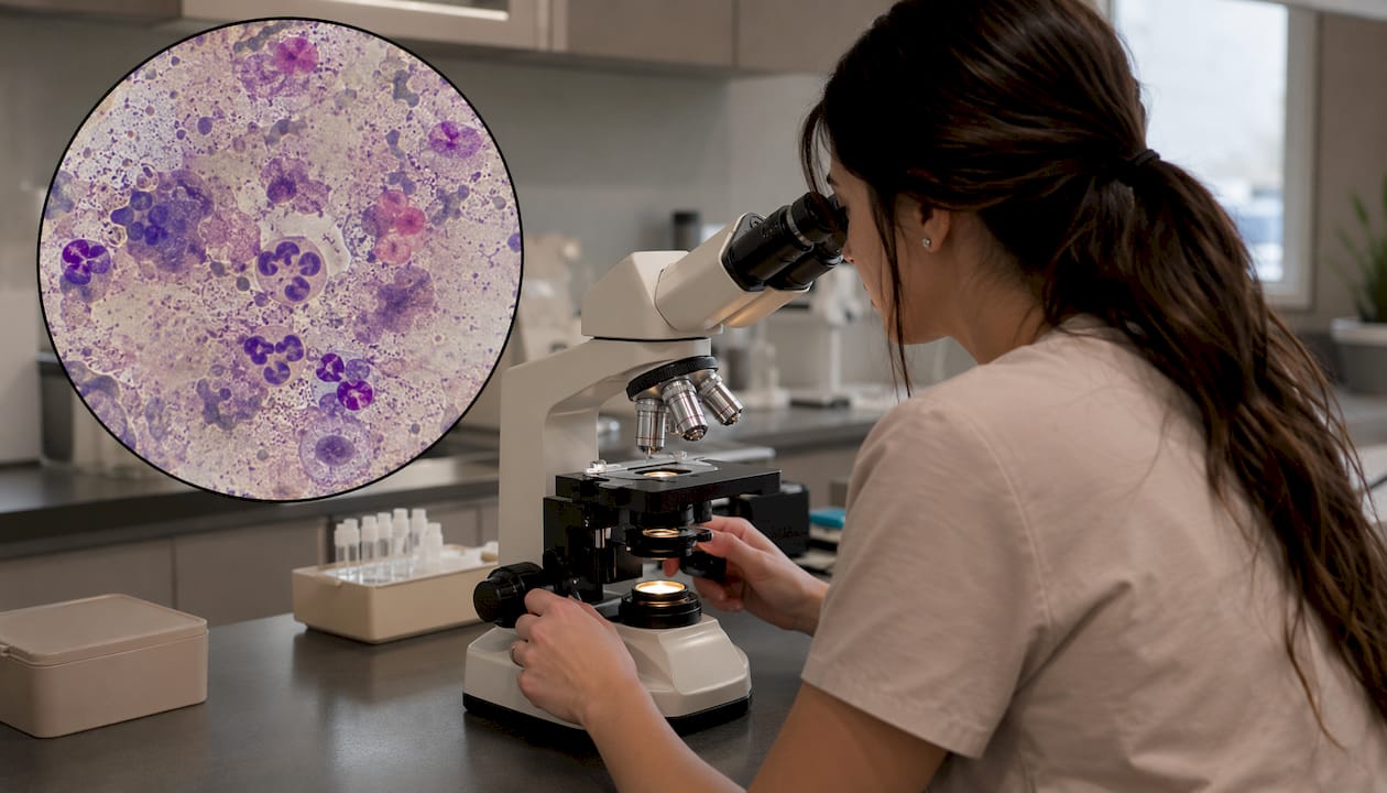
The single most important diagnostic move in canine cytology is distinguishing septic from sterile inflammation. Neutrophils with intracellular bacteria confirm septic pyoderma and mandate antimicrobial therapy. Neutrophils without organisms suggest sterile inflammation or early infection where organism load is low. Always correlate with clinical signs and prior therapy before concluding sterile.
Cell types and their clinical significance
| Cell type | Likely causes | Clinical clues |
|---|---|---|
| Degenerate neutrophils + intracellular bacteria | Septic pyoderma, septic otitis | Karyolysis, nuclear streaming; bacteria visible inside cells |
| Non-degenerate neutrophils, no organisms | Sterile pustular disease, immune-mediated | Acantholytic keratinocytes suggest pemphigus foliaceus |
| Eosinophils | Allergic/hypersensitivity reaction, parasitic, eosinophilic granuloma | Often concurrent with mast cells; check for ectoparasites |
| Mast cells | Mast cell tumor, hypersensitivity | Purple cytoplasmic granules; numerous mast cells warrant biopsy |
| Macrophages | Chronic inflammation, fungal infection, foreign body | Large vacuolated cells; look for intracellular organisms |
| Lymphocytes | Chronic inflammation, lymphoma | Small lymphocytes are reactive; large atypical lymphocytes raise neoplasia concern |
Artifacts and pitfalls. Nuclear streaming (smeared nuclear material between cells) results from excessive pressure during slide preparation and can mimic extracellular bacteria at low power. Crushed cells from aggressive smearing lose nuclear detail entirely. Prior topical therapy, particularly glucocorticoids or antibiotics, can dramatically reduce inflammatory cell numbers and organism counts, producing a falsely reassuring slide. Always ask about recent treatment before interpreting a "clean" result.
Normal canine skin hosts commensal organisms, including low numbers of Malassezia and cocci. The diagnostic move is to correlate organism density with inflammatory cells and clinical signs, not to treat organisms in isolation. A few cocci on a slide from a non-inflamed skin fold is not pyoderma.
Pro Tip: When a slide shows neutrophils but no organisms, ask yourself three questions before concluding sterile: Was the sample collected from the right site? Has the patient received antibiotics in the past two weeks? Is the organism load simply below the detection threshold of light microscopy? If any answer is uncertain, collect a second sample from a fresh lesion.
Recognizing bacteria, Malassezia, fungi, and ectoparasites on cytology
Morphologic pattern recognition is the core skill here. The key distinctions are cocci versus rods, intracellular versus extracellular location, and yeast morphology.
- Cocci appear as small round purple dots, often in clusters or chains. Staphylococcus pseudintermedius is the most common canine skin pathogen and presents as cocci. Intracellular cocci inside neutrophils confirm active infection rather than colonization.
- Rods (bacilli) are elongated, often larger than cocci, and may appear singly or in pairs. Rods on cytology are a hard escalation trigger: culture and sensitivity testing is mandatory because Gram-negative rods (Pseudomonas, Proteus, E. coli) carry a high probability of multidrug resistance.
- Malassezia yeast appear as peanut- or footprint-shaped organisms, typically 3–8 µm, staining blue-purple with Diff-Quik. Abundant yeast with minimal inflammation suggests overgrowth or dysbiosis. Yeast with concurrent neutrophils indicates true Malassezia otitis or dermatitis requiring antifungal therapy.
- Dermatophyte hyphae and spores are occasionally visible on tape prep from scaling lesions but are more reliably detected by Wood's lamp, fungal culture, or PCR. Cytology alone is not sufficient to rule out dermatophytosis.
- Demodex mites are best found on deep skin scrapings, but tape impression while squeezing the skin is a sensitive alternative that often detects more adult and larval mites than scraping alone. Mites appear as elongated cigar-shaped organisms at low power.
When to escalate to culture: rods on cytology, recurrent infection despite appropriate therapy, unusual organism morphology, suspected otitis media, organisms persisting after two full treatment courses, or any systemic signs suggesting bacteremia. Cytology guides targeted therapy and supports antimicrobial stewardship by identifying mixed yeast-bacterial dysbiosis before broad-spectrum drugs are prescribed.
Annotated photomicrographs are invaluable for training technicians and calibrating interpretation. Standard pictorial resources such as the Clinicians Brief cytology image library and the BSAVA Manual of Canine and Feline Dermatology provide representative images with scale bars for common organisms.
Common cytologic patterns and what they mean for treatment
Four patterns account for the majority of canine skin cytology findings in general practice.
Pyoderma. Degenerate neutrophils with intracellular cocci. This is the clearest cytologic diagnosis in dermatology. Systemic antibiotics are indicated; topical therapy alone is appropriate only for surface or superficial pyoderma with minimal lesion depth. Culture is not necessary for a first episode with cocci, but becomes mandatory if the infection recurs or rods appear.
Malassezia dermatitis. Numerous peanut-shaped yeast, often with variable neutrophilic inflammation. Topical azole therapy (miconazole, ketoconazole shampoo) is first-line for localized disease; systemic azoles (ketoconazole, itraconazole) are needed for generalized or recurrent cases. Always investigate the underlying cause, most commonly atopy or food allergy, because Malassezia dermatitis rarely resolves permanently without addressing the predisposing disease.
Superficial bacterial folliculitis. Intracellular cocci within follicular epithelial cells or within intact pustules. The cytologic picture is similar to surface pyoderma but the clinical context differs: folliculitis typically presents as papules and pustules in hair follicle distribution. Systemic antibiotics for 3–4 weeks minimum; recheck cytology at the end of treatment to confirm resolution.
Sterile pustular disease. Non-degenerate neutrophils without organisms, often with acantholytic keratinocytes. This pattern raises pemphigus foliaceus as the primary differential. Anti-inflammatory or immunosuppressive therapy replaces antibiotics here, which is exactly why cytology before treatment matters. A dog with pemphigus foliaceus treated empirically with antibiotics will not improve, and the delay costs weeks.
Short case examples: a 4-year-old Labrador with recurrent interdigital swelling showed rods on ear cytology at a recheck, prompting culture that confirmed Pseudomonas with a narrow sensitivity profile. Switching to a targeted fluoroquinolone resolved the infection within three weeks. In a separate case, a 7-year-old Golden Retriever with generalized scaling had abundant Malassezia on tape prep with no neutrophils; azole shampoo twice weekly cleared the lesions, and subsequent allergy workup identified a food component as the driver.
Pitfalls and when to escalate beyond cytology
Cytology is powerful but not infallible. Knowing its limits prevents mismanagement.
Common pitfalls:
- Misreading commensals as infection. Low organism counts without inflammation are normal. Commensal organisms exist in low numbers on healthy canine skin; diagnosis requires correlation of density with inflammatory cells and clinical signs.
- Sampling error. A slide from the wrong site, a poorly collected swab, or a lesion that has been treated topically can all produce a falsely negative result.
- Staining artifacts. Nuclear streaming, overstaining, and mineral deposits from tap water rinse can all mimic organisms or obscure cells.
- Low organism load in sensitized patients. A dog with a strong inflammatory response may have very few organisms visible despite active infection, because neutrophils are consuming them faster than they replicate.
- Biofilm-protected organisms. Biofilms, ear canal stenosis, and underlying allergic or anatomic causes can allow organisms to persist despite low counts on cytology. A "clean" slide does not guarantee cure.
Escalation criteria checklist:
- Rods visible on cytology at any count.
- Organisms persist on cytology after two appropriate treatment courses.
- Chronic or recurrent lesions without a clear underlying diagnosis.
- Nodules, ulcerative lesions, or draining tracts where neoplasia is possible.
- Systemic signs (fever, lymphadenopathy, weight loss) accompanying skin disease.
- Suspected otitis media (head tilt, Horner's syndrome, neurologic signs).
- Any lesion where the cytologic pattern does not match the clinical presentation.
Biofilm is a particular challenge in chronic otitis. Organisms embedded in biofilm are protected from both topical antimicrobials and immune cells, meaning organism counts on cytology may be low while clinical disease remains severe. Ear canal stenosis compounds this by limiting drug penetration. These cases need culture, sensitivity testing, and often referral to a veterinary dermatologist.
Practical in-clinic workflow: cost, turnaround, and talking to owners
In-house cytology costs a fraction of a culture submission and delivers results during the same appointment. The equipment investment is a light microscope with 10x, 40x, and 100x oil immersion objectives, Diff-Quik stain, and glass slides. Most practices already have this. Turnaround from sample collection to interpretation is typically 10–15 minutes.
Build cytology into the appointment flow for every otitis and skin lesion case. Collect the sample while the technician is taking history, stain while the veterinarian examines the patient, and read the slide before the treatment discussion. This sequence adds minimal time and eliminates the delay between diagnosis and therapy.
For documentation, use the reporting template from the microscopy section above. Record specimen type, collection technique, slide quality, semiquantified findings, interpretation, and next steps. This creates a clear audit trail for rechecks and referrals.
Owner communication does not need to be complex. A two-sentence script works well:
This framing sets expectations for the recheck, explains why treatment was chosen, and gives the owner a concrete timeline, all without requiring them to understand cytology.
Minimizing stress during sampling: Fear Free and home-visit adaptations
Sample quality depends partly on patient cooperation. A dog that is struggling or trembling produces smeared, artifact-laden slides. Reducing stress during the sampling sequence is both a welfare goal and a diagnostic one.
Low-stress sampling checklist:
- Ask owners to withhold food for two hours before the visit so high-value treats remain motivating during the appointment.
- Apply a dog-appeasing pheromone (Adaptil) spray to the exam table 15 minutes before the patient enters the room.
- Position the patient in lateral or sternal recumbency with a trained technician providing gentle restraint, rather than scruffing or forceful holds.
- Collect the most stressful sample (ear swab) last, after the patient has been rewarded for tolerating less invasive handling.
- Keep the sampling sequence short: collect, label, and release the patient to a comfortable position before staining and reading.
- Use a second technician for ear cytology in anxious dogs to allow one person to focus entirely on patient comfort.
Home-visit logistics. For severely anxious patients or owners who cannot transport their pets, a house call eliminates the travel and waiting room stress that often accounts for the worst behavioral responses. A portable cytology kit for home visits should include pre-labeled glass slides, acetate tape strips, cotton swabs, sterile needles, a Diff-Quik travel kit (individual dip containers), immersion oil, and a coverslip supply. Slides collected at home can be stained and read at the clinic within the same visit window if a battery-powered field microscope is available, or transported back for same-day reading.
City Animal Veterinary Center specializes in a unique Fear Free approach that minimizes stress for pets during visits, enhancing comfort for both pets and owners. City Animal Veterinary Center also offers a home visit service for clients who have difficulty traveling with their pets, emphasizing convenience and accessibility. These two service models make routine cytology achievable for patients who would otherwise go undiagnosed or undertreated due to handling challenges.
Pro Tip: For dogs with severe ear sensitivity, apply a small amount of lidocaine gel to the swab tip and wait 60 seconds before sampling. The swab goes in more smoothly, the dog tolerates it better, and the sample quality is often superior because the dog is not pulling away mid-collection.
Key Takeaways
Skin cytology in dogs is the fastest path from clinical suspicion to targeted therapy: collect by lesion type, stain with Diff-Quik, and interpret cells plus organisms together before treating.
| Point | Details |
|---|---|
| Match technique to lesion | Use impression smears for moist lesions, tape prep for dry/scaly skin, and swabs for ear canals. |
| Heat-fix selectively | Heat-fix dry or greasy smears only; never heat-fix moist samples or tape preparations. |
| Correlate, don't count alone | Counts above 5 yeasts or 25 bacteria per 40x field often suggest infection, but diagnosis must be confirmed with clinical signs. |
| Escalate on rods | Rods on cytology always require culture and sensitivity testing before treatment. |
| Cityanimalvet's approach | Cityanimalvet offers in-house cytology with Fear Free handling and home-visit diagnostics for anxious or immobile patients in Brooklyn, NY. |
Why routine cytology changed how I practice dermatology
The conventional wisdom in general practice used to be: skin infection, reach for a broad-spectrum antibiotic. Cytology has made that approach feel like guessing in the dark. Once you start reading slides routinely, the cases that would have gotten a two-week course of amoxicillin-clavulanate start sorting themselves out clearly: this one has rods and needs a culture, that one has nothing but acantholytic keratinocytes and needs a dermatologist, the third one has cocci inside neutrophils and will respond to cephalexin.
What surprises most clinicians is how often the slide changes the plan. Sterile pustular disease looks identical to pyoderma on physical exam. Malassezia dermatitis in a dog with concurrent bacterial infection gets missed if you only treat the bacteria. The slide tells you both are there. That is not a subtle refinement; it is the difference between a dog that improves and one that comes back in three weeks looking the same.
The stewardship argument matters too. Cytology reduces empirical use of broad-spectrum medications by identifying mixed dysbiosis and guiding targeted therapy. Every time a slide shows cocci and you prescribe a narrow-spectrum drug instead of a fluoroquinolone, you are preserving that fluoroquinolone for the case that actually needs it.
Cityanimalvet's dermatology services: what to expect at your appointment
Skin and ear problems are among the most common reasons dogs visit the vet, and getting the diagnosis right on the first visit saves time, money, and the dog's comfort. At Cityanimalvet in Brooklyn, NY, in-house cytology is part of the standard dermatology workup: samples are collected, stained, and read during your appointment so the treatment plan is based on what is actually on the slide, not a best guess.

The clinic's Fear Free-certified team uses low-stress handling techniques throughout the sampling process, which means better samples and a calmer dog. For patients who do not travel well or owners with mobility constraints, house call appointments bring the same diagnostic capability to your home. You do not need to bring anything special; just note any treatments the dog has received in the past two to three weeks, since recent antibiotics or antifungals affect what the slide shows.
To book a dermatology appointment or schedule a house call, visit Cityanimalvet or call the clinic directly. Same-day appointments are often available for acute skin and ear concerns.
Useful sources and further reading
The following resources form the clinical backbone of this guide and are recommended for clinicians who want deeper protocol detail or image libraries.
- Skin Cytology in Dogs and Cats (Canadian Veterinary Medical Association, 2019). Practical sampling and staining protocols including heat-fix guidance and technique selection by lesion type. Useful as a printable clinic reference.
- Ear Cytology: A Key Test in the Diagnosis and Management of Canine Otitis Externa (Veterinary Medicine Journal, 2022). Covers semiquantitative thresholds, battleship scanning technique, and the limitations of clean cytology in biofilm cases. Strong on escalation criteria.
- Ear Cytology in Otitis Externa: When, Why, How (The Veterinary Nurse). Technician-focused guide to sampling, staining, and antimicrobial stewardship rationale. Practical for training veterinary nurses.
- Skin Cytology: Dermatologic Disease in Dogs (Clinicians Brief). Comparative technique data on plain glass versus adhesive slides; includes photomicrographs of common findings. Recommended for image-based learning.
- The Normal Canine Skin Microbiome and Clinical Implications (PMC). Peer-reviewed data on commensal organism populations; essential context for avoiding overdiagnosis of infection from low organism counts.
- Chronic Otitis in Cats: Clinical Management (PMC). Covers cat ear cytology reference intervals, Malassezia prevalence, and the stain artifact from feline cerumen. Useful for practices seeing both species.
- BSAVA Manual of Canine and Feline Dermatology. The standard pictorial reference for cytologic patterns, photomicrograph atlases, and biopsy correlation. Recommended for any practice doing regular dermatology work.
- Mucky Ears, Swabs and Smears: A Guide to Interpreting Diagnostics for Otitis Externa (BSAVA/IDEXX, 2025). Up-to-date practical guide covering impression smears, tape prep, and Diff-Quik staining for otitis cases; supports the "every otitis patient" recommendation.