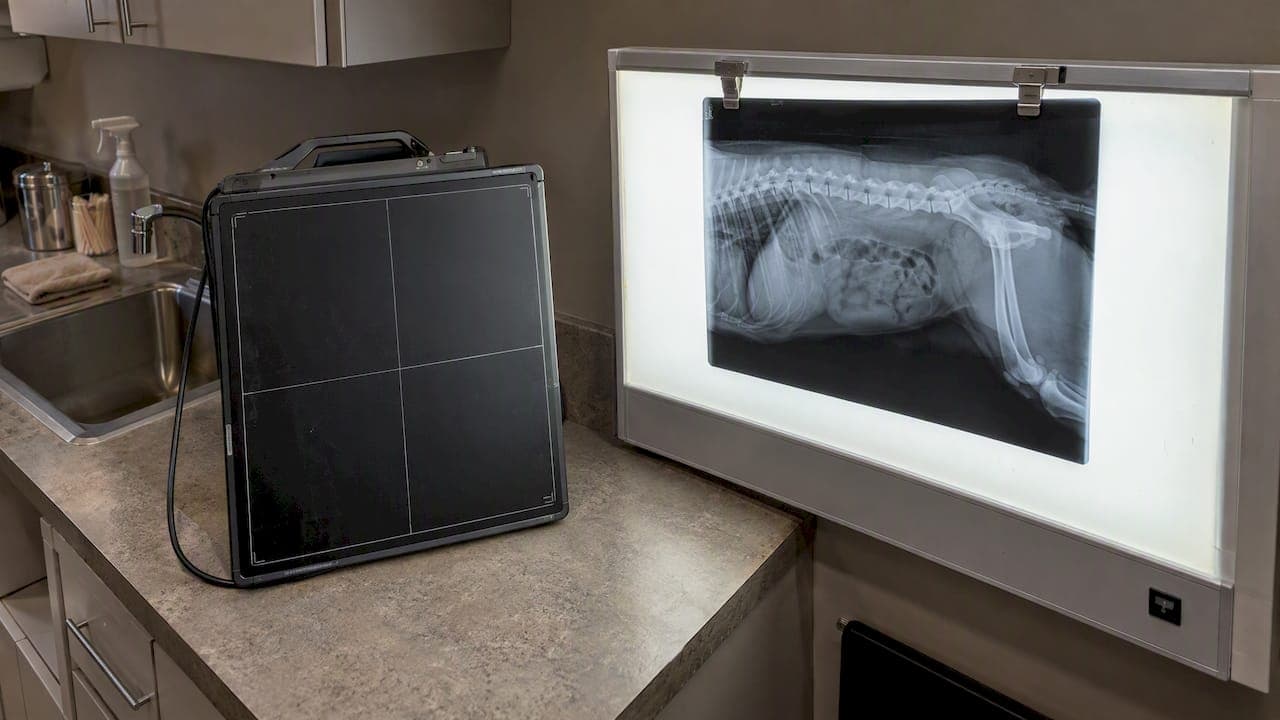
For most routine imaging, digital X-rays produce equal or better diagnostic images than film while allowing a lower routine dose and a faster workflow. That holds as long as the clinic watches its exposure indices to prevent dose creep. Digital also lets us review, adjust and share images quickly. Here is what that means for your pet and for the team behind the camera.
TL;DR:
- Digital detectors can hide overexposure, so clinics should review exposure indices on every image and use scheduled quality checks to prevent dose creep.
- For fine bone detail, film historically resolved more line pairs, but modern direct digital detectors meet routine clinical needs; computed radiography plates fall between them.
- Digital images appear immediately, letting clinicians check image quality before the pet leaves the table; positioning errors can still require repeat exposures.
- Digital eliminates film chemicals and physical archives, but clinics take on detector costs, software support, backups, cybersecurity, and eventual equipment replacement.
Table of Contents
- Digital X-ray vs film at a glance
- How does image quality compare in real practice?
- Radiation dose: what the evidence says and how clinics prevent dose creep
- How does digital change workflow, retakes and storage?
- What operational and environmental differences should clinics weigh?
- How do the costs of digital and film X-rays compare?
- What are the maintenance, repair and durability differences?
- Does digital imaging change diagnostic accuracy in veterinary medicine?
- How do digital X-rays work with medical records and telemedicine?
- What training do veterinary staff need for digital X-rays?
- Our perspective: low-stress, safer imaging with digital X-rays
- Get a calm, quick X-ray for your pet at City Animal
- FAQ
- Sources
Digital X-ray vs film at a glance
- Image quality: Digital detectors capture a wider range of tissue density in one exposure, and the image can be brightened, contrasted and magnified on screen. Film is fixed once it is developed.
- Radiation: Digital systems can need less dose per image when protocols are tuned, but they also hide overexposure, so quality assurance matters more, not less.
- Workflow: Digital images appear almost immediately, retakes are less common, and sharing with a specialist takes a click instead of a courier.
- Environment and operations: Film means developer, fixer and physical archives. Digital removes the chemicals and replaces them with IT, backups and software updates.
- Diagnostic confidence: Zooming and windowing a digital image often reveals detail that a single fixed film exposure cannot, without exposing your pet again.
How does image quality compare in real practice?
"Better image" means several things at once: how many shades of gray the system can show, how much contrast you can pull out of a difficult exposure, and how fine a detail it can resolve.
Dynamic range is where digital pulls ahead most clearly. A peer-reviewed comparison of digital radiography with conventional imaging describes digital radiography as offering a wider dynamic range than screen-film, along with reusable plates or solid-state detectors and immediate image availability. In practical terms, a chest film exposed so the lungs look right often leaves the heart and spine too white to read. A digital image can show both regions, and the clinician can adjust the window and level on screen to examine each one.
Post-processing is the second advantage. Digital radiographs can be magnified, contrast-adjusted and brightness-modified without re-exposing the patient. With film, a slightly dark or light image means either squinting at it or taking another picture. For a nervous cat or a dog in pain, avoiding a retake is a real welfare benefit, not a minor convenience; understanding dog dental health nutrition also plays a key role in overall pet well-being.
Spatial resolution is the point film loyalists raise, and it deserves a fair answer. Film historically resolved very fine line pairs, and in a few very-high-detail situations, such as subtle bone texture changes in small joints, that edge was real. The same review notes that solid-state flat-panel detectors deliver better quality with less radiation than screen-film radiography, so modern direct digital detectors meet clinical needs in nearly every routine case. Computed radiography plates, the older digital approach, sit somewhere between the two.
So when do the small resolution differences matter? Mostly in fine musculoskeletal detail, where a clinician may want every possible line pair. When do post-processing and dynamic range decide the case? Far more often: thoracic imaging, abdominal studies, and any patient whose thickness varies across the field of view. In everyday practice, an image you can adjust beats a slightly sharper image you cannot.

Radiation dose: what the evidence says and how clinics prevent dose creep
Digital detectors can allow a lower dose to produce a diagnostic image, but only when the protocol is set up that way. The ACR–AAPM–SIIM–SPR practice parameter for digital radiography directs that digital radiography deliver the necessary image quality at an appropriate radiation dose, and it stresses that qualified personnel and quality assurance programs are essential. In other words, the technology makes lower dose possible. People and process make it happen.
The risk that comes with that flexibility is called dose creep. Film punishes overexposure immediately: the image turns black and has to be repeated. Digital detectors have such wide latitude that an overexposed image still looks fine, because the software rescales it. A published study on dose creep in digital radiography identifies it as a critical operational risk precisely because images can look acceptable despite higher exposures, which means increases can go unrecognized without active monitoring.
Digital detectors can mask overexposure, so exposure indices must be monitored actively rather than judged by how the image looks.
Phantom research points the same way. A study of dose creep potential in computed radiography found a high tolerance for overexposure, with perceptual image quality staying acceptable across a wide exposure range. Professional commentary in the radiography literature makes the same point: digital detectors can hide overexposure, so monitoring exposure indices and running a quality control program keeps doses as low as reasonably achievable.
Here is what a well-run clinic does to stay on the right side of that line:
- Checks the exposure index on every image, not just the ones that look wrong.
- Builds species- and size-specific technique charts and reviews them on a schedule.
- Runs routine quality assurance checks on detectors and generators.
- Reviews exposure trends across technicians, so drift is caught early.
- Keeps oversight shared between clinicians, technicians and, where available, a physicist.
For owners, the takeaway is simple: ask whether your clinic tracks exposure indices. A good answer is a sign of a safety culture, not just a nice piece of equipment.
How does digital change workflow, retakes and storage?
The change owners notice first is speed. A digital image appears quickly, so the clinician can confirm positioning and quality before your pet leaves the table. With film, you wait for processing, and a bad exposure means going back and doing it again.
- Faster visits: Immediate image availability shortens the imaging portion of an appointment, which matters a great deal for animals that are anxious or uncomfortable.
- Fewer retakes: Because exposure latitude is wide and the image can be adjusted on screen, fewer studies need to be repeated for exposure errors. Positioning errors still happen, but they are caught right away.
- Easy storage and retrieval: Images live on a network archive, often called PACS, rather than in paper envelopes. The comparison review lists network storage as one of digital radiography's core advantages.
- Simpler second opinions: Sending a study to a specialist or a referral hospital takes a link or a file transfer, not a courier or a photocopy of a fragile film.
All of this adds up to better throughput for the clinic and a calmer experience for you. Less time on the table, fewer repeat exposures, and quicker answers are benefits you feel in the room, even if you never see the technology behind them.
What operational and environmental differences should clinics weigh?
Film comes with a physical footprint. It needs a darkroom or automatic processor, developer and fixer chemicals that have to be handled and disposed of properly, and shelves or filing rooms to store the results. Films also degrade with time, so an old study may be harder to read than it used to be.
Digital removes the chemistry and the paper archive, which is a clear environmental gain. In exchange, it adds a different set of responsibilities: computers and servers, backup systems, cybersecurity, software licenses and periodic updates. A clinic that adopts digital takes on IT as part of its core operations.
Lifecycle thinking is useful here. Digital detectors and software eventually need replacing or upgrading, while film practices need a continuing supply of film, chemicals and processor maintenance. Neither route is effortless. The practical question is which set of tasks a clinic can run reliably, and most practices find that managing servers and backups is easier than managing chemical handling and long-term film storage.
How do the costs of digital and film X-rays compare?
We leave dollar figures to the clinics that quote them, but the structure of the comparison is worth understanding. Film is a pay-as-you-go system: every image uses film and processing supplies, and the equipment needed to start is relatively modest. Digital flips that pattern. The detector and software are the larger commitment up front, and each additional image costs very little to produce.
Over the long run, digital also avoids recurring spending tied to chemistry, film stock, disposal and physical storage space. It adds other ongoing items, such as software support, data backup and eventual detector replacement. The honest summary is that the two systems spend money in different places, and the right choice depends on how many studies a clinic performs and how it plans to maintain its equipment.
For pet owners, the more useful angle is value in the exam room: fewer repeat exposures, faster answers and easier sharing. Those benefits show up in your pet's experience regardless of how a clinic structures its budget.
What are the maintenance, repair and durability differences?
Film systems depend on mechanical and chemical parts: processors with rollers and heaters, chemistry that must be replenished and monitored, and cassettes and screens that wear and collect scratches or dust, which show up as artifacts on the image. When a processor fails, imaging stops until it is fixed.
Digital systems shift the maintenance burden. Solid-state flat-panel detectors have no chemistry and few moving parts, but they are precision electronics, so they need careful handling, protection from drops and scheduled calibration. Computed radiography plates are reusable but still wear with handling. Software needs updating, and the network and backup systems that hold your images need regular checks.
The ACR practice parameter emphasizes technical standards and qualified oversight for digital workflows, which is another way of saying that maintenance is not optional. A detector that drifts out of calibration can degrade quality or quietly alter dose. Clinics that treat quality assurance as routine, with scheduled checks and service agreements, keep digital equipment reliable for years. Film equipment can also last a long time, but it never stops depending on consumables and chemistry.

Does digital imaging change diagnostic accuracy in veterinary medicine?
In practice, the biggest effect on diagnosis comes from what a clinician can do with the image. Animals vary enormously in size and body shape, and a fixed exposure rarely suits every tissue in the field. Digital's wide dynamic range and on-screen adjustments make it easier to see subtle lung patterns, small foreign objects in the gut, or fine changes along a bone edge without repeating the exposure.
Magnification and contrast control also help with common veterinary questions: Is that a fracture line or an overlapping shadow? Is there fluid in the chest? Is the heart silhouette normal? Being able to window an image and compare regions turns a borderline picture into a clearer answer more often than a fixed film does.
There is also a patient benefit that is easy to overlook. When the clinician can confirm quality immediately, fewer animals face repeat exposures and extra restraint. For a pet with a painful limb or a stressed airway, that matters. We would stop short of claiming that digital changes every outcome, and the comparison literature is mostly about image performance and dose rather than veterinary survival data. What it consistently shows is better tools for the person reading the image, and that is where diagnostic confidence comes from.
How do digital X-rays work with medical records and telemedicine?
Digital images are files, which makes integration the quiet strength of the format. A radiograph can be attached directly to your pet's electronic medical record, sitting beside the exam notes, lab results and history, so the whole picture is in one place. Nobody has to hunt for an envelope or reconcile a paper film with a chart.
Telemedicine and referrals benefit just as much. When a case needs a board-certified radiologist or a surgical specialist, the study can be sent electronically, and the specialist views the same adjustable image the clinic saw. The comparison review lists network storage and ready sharing among the practical advantages of digital radiography, and for referrals that translates directly into faster answers for owners.
Compatibility does take some care. Clinics need imaging software that works with their records system, sensible file naming, secure transfer methods and reliable backups. Done well, it also means your pet's imaging history follows them: if you move, change practices or see a specialist, prior studies can be compared with new ones, which is one of the most useful things a radiograph can do.
What training do veterinary staff need for digital X-rays?
Moving to digital does not remove the need for skill. It changes which skills matter most. Positioning, collimation and technique selection remain the foundation of a good radiograph, and no software can fix a poorly positioned limb.
What staff add is a new layer of knowledge: understanding exposure indices, recognizing that an image can look fine and still be overexposed, and knowing how to use post-processing responsibly rather than relying on it to rescue poor technique. The ACR practice parameter stresses the qualifications and responsibilities of personnel, as well as technical standards for digital workflows, which reflects how central training is to getting the dose benefits digital promises.
Good training programs cover several areas: the basics of how the detector captures and processes an image, how to read and act on exposure indices, how to use technique charts for different species and sizes, and how to run basic quality checks. Ongoing review matters as well, because dose creep develops gradually. A team that looks at its exposure data regularly and talks openly about it will keep doses low long after the initial training fades.
Our perspective: low-stress, safer imaging with digital X-rays
We use digital X-rays for diagnostic imaging, and the choice goes hand in hand with how we run our Fear Free approach. Because images appear immediately, we can confirm quality quickly and keep imaging time short, which means less time on the table for an anxious pet. We use a Fear Free approach to imaging, and you can read more in our guide to pet X-ray safety for owners. We also aim to make imaging a calm experience for pets. For a sense of how we approach calm imaging more broadly, see our Fear Free ultrasound prep guide.
— CAVC
Get a calm, quick X-ray for your pet at City Animal
If your pet needs an X-ray, we offer digital imaging in our clinic with a Fear Free approach, and house calls when they are appropriate. We use a Fear Free approach and can talk through the results with you.

You can learn more about our diagnostic imaging service, or ask about our home visit options. Reach out through either page to book or ask a question.
This article is general information, not a substitute for advice from a qualified doctor. Consult a qualified healthcare professional about your own circumstances before acting on anything here.
FAQ
Why is digital radiography better than film?
Digital radiography offers a wider dynamic range, immediate image availability and on-screen adjustment without re-exposing the patient. It also supports network storage and easy sharing. Those advantages are described in a peer-reviewed comparison of digital and conventional imaging, and they add up to faster, more flexible diagnosis.
What are the downsides of digital radiography?
The main downside is dose creep: wide detector latitude can make overexposed images look acceptable, so clinics need to monitor exposure indices and run quality assurance. Digital systems also bring IT, backup and software maintenance tasks that film did not require.
Are all X-rays digital now?
Digital has become the standard approach in many practices, but film and older workflows can still turn up, particularly in historical records and in some niche or legacy setups. Clinics that have moved to digital typically keep archives of older film studies for comparison.
What are three advantages of digital X-rays over film X-rays?
First, a wider dynamic range and on-screen adjustment give clinicians more diagnostic information from a single exposure. Second, images are available almost immediately, which shortens visits and reduces retakes. Third, electronic storage makes sharing with specialists and attaching images to records much simpler.
Sources
- Digital radiography. A comparison with modern conventional imaging - PMC
- ACR–AAPM–SIIM–SPR PRACTICE PARAMETER FOR DIGITAL RADIOGRAPHY
- Researchprofiles