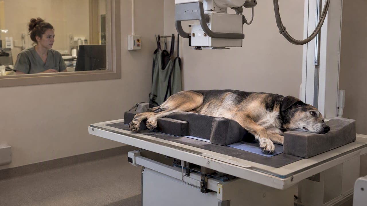
Routine veterinary X-rays are generally safe for pets when performed with standard radiation-safety measures. Clinics follow the ALARA principle (as low as reasonably achievable) and rely on shielding, positioning aids, and hands-free techniques rather than holding pets during exposure. You typically wait outside the room while images are taken, and if sedation is needed to get a clear picture, your veterinary team follows standard pre-anesthetic safety steps first.
TL;DR:
- Ask whether the clinic uses hands free positioning by default, since sandbags and supports keep staff out of the beam during exposure.
- Sedation may suit pets that are painful, anxious, or need a precise position; expect an exam and bloodwork beforehand, plus monitoring during recovery.
- For a pregnant pet, discuss necessity and timing with the veterinarian; X rays are generally limited to late gestation, and ultrasound uses no ionizing radiation.
- Clear positioning avoids retakes and extra exposure; ask how many images are expected and whether fasting is needed if sedation is planned.
Table of Contents
- How X-rays work and why veterinarians use them
- How much radiation exposure is really involved
- How clinics keep X-rays safe for pets and staff
- When vets choose sedation over physical restraint
- Special situations: pregnancy, young pets, and repeat imaging
- How to prepare your pet and what to ask before X-rays
- Our Fear Free approach to imaging at City Animal Veterinary Center
- Book a low-stress imaging visit with our team
- FAQ
- Sources
How X-rays work and why veterinarians use them
A radiograph is a brief, targeted burst of ionizing radiation that passes through tissue to create an image on a detector. Dense structures like bone absorb more radiation and appear white, while soft tissue and air show up in shades of gray. According to the Weill School of Veterinary Medicine, radiographs are a fast, common, and typically painless diagnostic tool used across small animal practice.
Veterinarians order X-rays for a wide variety of reasons, and the exposure is justified by the diagnostic value:
- Suspected fractures or joint injuries after trauma
- Gastrointestinal obstructions, such as a swallowed foreign object
- Heart and lung evaluation for coughing, breathing changes, or murmurs
- Dental radiographs to check roots and bone below the gumline
Good positioning matters as much as the equipment itself. A pet that moves, or is positioned incorrectly, often produces a blurry or incomplete image that has to be repeated, which means more handling and more exposure than a single clean shot would require.
How much radiation exposure is really involved
A single diagnostic radiograph delivers a small, brief dose, and the bigger safety question in most clinics is staff exposure over repeated procedures rather than risk to any one patient. The ACVR radiation safety statement endorses the ALARA principle: minimize time near the beam, maximize distance from it, and use shielding whenever possible.

Staff doses stay low when protocols are followed, though they can rise with larger patients, certain technique settings, or manual restraint, according to a real-time dosimetry study of veterinary radiography procedures. That finding is part of why the same research supports avoiding manual restraint whenever a mechanical alternative exists.
This is also why many practices use personal dosimeters to track cumulative occupational exposure for the people taking the images, not just the animals being imaged. A single study on a dog or cat is a minor, well-justified exposure; the safety systems in place exist mainly to protect the humans in the room day after day.
How clinics keep X-rays safe for pets and staff
The biggest shift in veterinary radiography over the past decade has been away from manually holding pets in position and toward hands-free techniques. Sandbags, foam troughs, positioning tape, and velcro straps now do the job that a technician's hands once did, which keeps people out of the direct beam.
- Hands-free positioning tools replace manual restraint for most routine views
- Portable units are used carefully, since they can expose more of the room to scatter radiation than a shielded, stationary unit
- Staff wear lead aprons, thyroid collars, and sometimes lead glasses, and they step behind a shielded barrier or leave the room during the actual exposure
- Technique settings (kilovoltage and milliamperage) are adjusted to the patient's size so one exposure captures a diagnostic image instead of requiring a retake
The ACVR points to hands-free initiatives, sometimes called Hands-Free X-Ray or Lower the Dose, as evidence that procedural standards across the profession are improving. A clinic that has adopted these habits is reducing unnecessary exposure for the people working there, not just following a checklist.
Pro Tip: Ask whether your clinic uses hands-free positioning as a default. A quick "yes, always" is a good sign of a well-run imaging protocol.
When vets choose sedation over physical restraint
Some pets need to be still for longer than a calm hold allows, and that is when sedation becomes the safer option rather than an extra step. Motion blur ruins an image, and a frightened or painful animal straining against a hand or a sandbag is both harder to image and more likely to get hurt in the process.
- Sedation is considered when a pet is in pain, anxious, aggressive, or needs a prolonged or precise position (such as for orthopedic views)
- A pre-anesthetic workup, including bloodwork and a physical exam, is standard before sedation to catch issues that would raise anesthetic risk
- Monitoring continues through recovery, not just during the procedure itself
- Vets weigh temperament, health status, and how critical the image is before deciding sedation is worth its own, separate risks
Pre-anesthetic workups and monitoring are standard practice specifically to reduce anesthetic risk, not an optional extra. The trade-off is straightforward: sedation adds a small procedural risk of its own but often reduces the number of retakes, the stress on the pet, and the physical struggle that manual restraint would otherwise require.
Special situations: pregnancy, young pets, and repeat imaging
Pregnant pets raise understandable concern, and the evidence here is reassuring but comes with caveats. A peer-reviewed review on imaging in the pregnant bitch found limited direct evidence of harm from a single radiograph, but it recommends limiting X-rays to late gestation when truly necessary, minimizing stress, and favoring physical restraint over chemical sedation unless there is a clear clinical reason to sedate.
- Discuss timing and necessity case by case with your veterinarian if your pet is pregnant
- Ask about ultrasound as an alternative, since it uses no ionizing radiation and is often preferred for monitoring pregnancy
- Avoid repeat imaging that isn't diagnostically necessary; good technique on the first attempt is the best way to limit cumulative exposure
How to prepare your pet and what to ask before X-rays
A little preparation on your end makes the visit smoother and often reduces how much handling or sedation your pet needs.
- Ask whether fasting is required; it usually only applies if sedation is planned
- Bring a comfort item, like a favorite blanket or toy, to ease waiting-room stress, following the kind of calming steps that help anxious dogs feel more at ease
- Ask directly: will anyone hold my pet, or is this a hands-free procedure?
- Ask what shielding and positioning aids will be used
- Ask whether sedation is planned, and why, so you understand the reasoning
- Ask how many images are expected, since a clear plan reduces the chance of surprise retakes
- Expect to wait outside the exposure room, and if sedation is used, expect a short recovery period with monitoring before you head home
Our Fear Free approach to imaging at City Animal Veterinary Center
We built our imaging workflow around reducing fear, not just radiation. For anxious cats, we may use pre-visit sedation with Bonqat at 5 mg/kg so a pet arrives already calm rather than fighting restraint in the exam room, and our ultrasound prep protocols follow the same Fear Free philosophy. When a pet truly cannot tolerate clinic travel, our home visit option lets us bring diagnostic imaging to a familiar space instead. When sedation is part of the plan, we follow informed anesthesia protocols with a pre-anesthetic workup and recovery monitoring.
— CAVC
Book a low-stress imaging visit with our team
If your pet needs an X-ray, the question isn't only whether the radiation is safe, it's whether the whole experience is handled with care. Our diagnostic imaging services pair hands-free technique with a Fear Free environment, so your pet is less likely to need retakes or heavier sedation just to hold still. Pets with dental concerns can get radiographs as part of our dental care visits, and when sedation is on the table, our team starts with the same pre-anesthetic groundwork described above.

For pets who struggle with clinic visits entirely, our home visit services bring certain diagnostic and wellness care directly to your door. Reach out to schedule an imaging appointment or ask our team which option fits your pet best.
This article is general information, not a substitute for advice from a qualified doctor. Consult a qualified healthcare professional about your own circumstances before acting on anything here.

FAQ
Do dogs have to be sedated for an X-ray?
Not always. Many dogs can be positioned with hands-free tools like sandbags and positioning troughs without any sedation, especially for routine chest or abdominal views. Sedation is reserved for painful, anxious, or uncooperative patients, or when a precise position must be held longer than a calm dog will tolerate.
What is the 15% rule in X-ray?
This refers to a radiography exposure-technique guideline used to adjust image contrast when changing kilovoltage, rather than a pet safety rule. It is a technical setting veterinary and medical radiographers use to fine-tune image quality, not a measure of radiation risk to patients.
What is the leading cause of death in dogs?
This is outside what our clinical imaging guidance covers, and claiming a specific ranked cause without a dedicated source would be misleading. Your veterinarian can speak to the main health risks for your dog's age, breed, and history during a wellness visit.
Do pets go through an X-ray machine at the airport?
No. According to TSA guidance, pets must be removed from their carriers at the checkpoint; the empty carrier goes through the X-ray machine while you carry or walk your pet through the metal detector. If you are flying with a pet, reviewing airline carrier and screening requirements ahead of time helps the process go smoothly.
Are there alternatives to sedation for anxious pets during X-rays?
Yes, non-pharmacologic calming strategies, patient conditioning, and gentle handling techniques can reduce the need for sedation in some pets, and non-drug anxiety management approaches are increasingly used alongside standard restraint methods. Whether these are enough depends on the individual pet's temperament and the type of image needed.
Sources
- ACVR radiation safety statement
- TSA offers tips for traveling with small pets through security checkpoint
- Radiography (X-ray) | Weill School of Veterinary Medicine
- Occupational radiation exposure of zoo veterinarians during x-ray procedures: a real-time dosimetry study