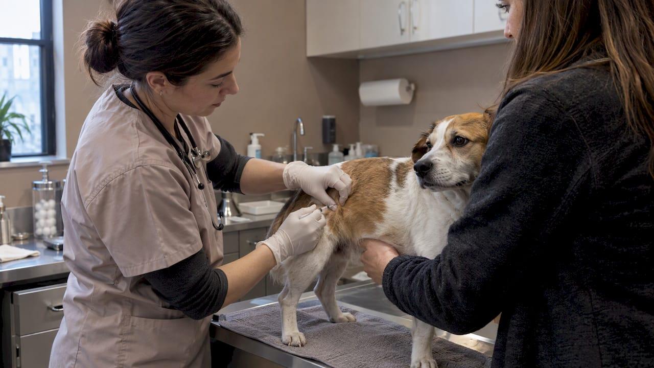
A fine needle aspirate (FNA) is a quick test in which a veterinarian uses a thin needle to collect a small sample of cells from a lump, then examines those cells under a microscope. For most dogs it is done during an ordinary exam, needs no sedation, and gives the first real answer to the question every owner asks when they find a lump: what is this? At City Animal Veterinary Center in Brooklyn, a lump is a reason to book an exam promptly, and our Fear Free approach is built to keep your dog calm while the team looks into it.
TL;DR
- An FNA collects cells from a lump or fluid with a thin needle so they can be read under a microscope. It is the most frequently used technique for collecting cells for cytology.
- It is minimally invasive and typically does not require general anesthesia or sedation. The sampling itself is brief.
- Results often come back within 0 to 2 days, and they can separate harmless lumps like fatty growths from tumors that need surgery.
- Sometimes the sample is inconclusive. That does not mean the test failed: a biopsy is the usual next step.
- A new lump deserves an exam sooner rather than later, because small tumors are easier to treat.
Table of Contents
- What a fine needle aspirate is, in plain terms
- When your vet is likely to recommend one
- How the procedure goes
- Calm matters more than you might think
- Reading the results
- Safety: what to know about risks
- After the result: the rest of the picture
- A checklist for the visit
- Have a lump you want checked?
- FAQ
- Sources
What a fine needle aspirate is, in plain terms
Your veterinarian cannot tell by touch what a lump is. A soft, movable bump can be a harmless fatty growth, and a similar-feeling one can be a tumor. The AAHA 2026 oncology guidelines say the type and likely behavior of a mass cannot be determined from palpation or imaging alone (AAHA is the American Animal Hospital Association, which sets practice standards for companion animal hospitals). Looking at the cells is how you get past guessing.
In an FNA, a small needle on a syringe is placed into the abnormal tissue, a few cells are collected, and they are spread on a glass slide. The slide is stained and read under a microscope. This cell-level study is called cytology. The University of Illinois College of Veterinary Medicine describes the aspirate as the usual first step in working up a pet's mass.
What can be sampled
Lumps under or on the skin are the classic target, but they are not the only one. PetMD lists masses in the mouth, masses attached to organs such as the spleen or liver, body fluids like joint fluid, and abnormal fluid in the abdomen or chest as common FNA uses. Lymph nodes are another frequent target: AAHA recommends sampling when spread to a lymph node is suspected, because a node's size and feel are not reliable indicators.
When your vet is likely to recommend one
Expect a recommendation when:
- You find a new lump, or an old lump grows, changes color or texture, or begins to bother your dog.
- A lump is found at a routine wellness exam.
- A lymph node feels enlarged.
- Ultrasound or X-rays show something unusual and the team needs to know what kind of cells are involved.
- There is fluid where there should not be any.
Timing matters. A veterinary oncologist quoted by DVM360 points out that most skin and subcutaneous (under-the-skin) tumors can be cured with surgery alone if they are found early, when they are small. That is the reason "wait and watch" is a poor plan for a lump nobody has sampled. If you have found one, an exam is the right first step. You can book a wellness exam and tell us about the lump when you call so we can plan the visit.
How the procedure goes
Before the visit
There is generally no special preparation. PetMD notes that most FNAs can be done during a routine appointment, with no special preparation or aftercare. It helps to write down when you first noticed the lump, whether it has changed in size, and whether your dog licks or scratches it. Photos taken next to a coin or ruler give the team a useful baseline.
During sampling
The standard needle is thin: Virginia Tech's veterinary protocol specifies a 22 to 25 gauge needle, three-quarters of an inch to an inch and a half long, with a small syringe and glass slides. The needle is similar in size to the ones used for many routine injections.
There are two common ways to collect cells. In aspiration, gentle suction is applied through the syringe. In the non-aspiration technique, sometimes called fenestration, the needle is moved through the lump without suction. Dr. Ed Branam, writing in Today's Veterinary Practice, notes that the non-aspiration technique often yields more cellular material and less bleeding, while aspiration suits fluid-filled masses. Your vet picks the approach based on the lump, and may take several passes in different spots so the sample represents the whole mass.

Does it hurt, and does my dog need sedation?
This is the question owners ask most. The needle prick is brief and most dogs tolerate it well. The Virginia Tech protocol describes the procedure as minimally invasive, one that does not typically require general anesthesia or sedation. Mild bleeding or bruising at the site is the most common after-effect, and it should fade over time.
Some situations are different. A very nervous or wiggly dog, or a lump in a sensitive spot, may be easier and safer to sample with light sedation. Fluid or masses deep inside the body are usually sampled with ultrasound guidance so the needle goes exactly where it should. Our guide to dog X-ray sedation explains how sedation decisions are made for diagnostic tests, which follows the same logic.
Calm matters more than you might think
A frightened dog moves, tenses and struggles, and that makes any sampling harder on the dog and the people. Anxiety is one of the main reasons owners put off a vet visit when they find a lump.
Fear Free is a certification program that trains veterinary teams in reducing fear, anxiety and stress during care. It is the approach we use at City Animal. In practice it means that the visit is paced to your dog, handling stays gentle, and the aim is for a dog to come back next time without dread. For an owner of an anxious dog, that can be the difference between a lump checked this week and a lump put off for months.
If getting your dog into a carrier, a car or a waiting room is the obstacle, ask us about home visits. Many dogs are more relaxed on their own couch, and it removes the logistics for busy professionals and for families with children and several pets.
Reading the results
How long they take
Cytology is fast. AAHA's 2026 guidelines give a turnaround of 0 to 2 days for cytology versus 3 to 7 days for histopathology, the tissue test that requires surgery or a tissue biopsy. Having an in-house laboratory at the practice helps keep the process moving. Slides that need a specialist's reading take a little longer, and your vet will tell you when to expect the answer.
What the cells can show
According to AAHA, FNA cytology often gives a definitive diagnosis of benign lesions such as a dermal cyst, a lipoma (a benign fatty lump) or inflammation, and of round cell tumors such as lymphoma, mast cell tumor and plasma cell tumor. The University of Illinois adds that round cell tumors and carcinomas are easily identifiable this way.
The result shapes the plan. PetMD gives the typical contrast: a mass of fat cells may be a benign lipoma that only needs monitoring, while a mass of mast cells may be a mast cell tumor with surgery planned to try to cure it. Most owners are relieved to learn how often the first answer is the benign one. When it is not, you have found out early, which is when options are widest.
Here is a quick way to think about the possible outcomes:
| Result | What it usually means | Typical next step |
|---|---|---|
| Benign (such as lipoma or cyst) | The lump is not cancer | Monitor, or remove if it bothers your dog |
| Tumor identified (such as mast cell tumor or lymphoma) | Cells point to a specific tumor type | Staging tests and a treatment plan |
| Inflammation or infection | The lump is a reaction, not a tumor | Treat the underlying cause |
| Inconclusive | Sample did not settle the question | Repeat sampling or biopsy |
When the sample is inconclusive
An inconclusive result is common enough that you should know about it in advance. PetMD says it can happen when a mass is very small, when the sampled spot does not represent the whole lesion, or when the lump does not release cells readily. In that case the next step is generally a surgical biopsy.
The University of Illinois makes the same point: if FNA does not give a diagnosis, a biopsy is often the next step. The AAHA summary published by DVM360 stresses that when the aspirate is inconclusive, or when a tumor grade is needed, clinicians should pursue biopsy and histopathology rather than assume how a tumor will behave. Biopsy tells more: it provides tumor grade and the width of surgical margins, which cytology cannot.
If surgery is the answer, whether to remove a lump or send tissue for histopathology, our team can walk you through it on the surgery page or at your exam.
Safety: what to know about risks
FNA is low risk, but a few points are worth knowing so you can watch your dog afterward:
- Bleeding and bruising are the most common effects. They should decrease over time, and infection is rare because the equipment is sterile.
- Mast cell tumors can swell after being touched or sampled, according to PetMD. Tell the veterinarian if a lump suddenly gets bigger, redder or itchier after the visit.
- Sampling inside the chest or abdomen carries a higher bleeding risk than a surface lump, which is why those samples are taken with imaging guidance and a plan.
- Bladder masses are handled differently. PetMD notes that aspirating a bladder transitional cell carcinoma can spread it into the abdomen, so urine sampling by catheter is recommended instead.
Call your veterinarian the same day if the site keeps bleeding, becomes hot, swollen or painful, or if your dog seems unwell, stops eating or is lethargic. If it happens outside hours, our urgent care page explains how to reach us.
After the result: the rest of the picture
Cytology answers "what are these cells?" If the answer is a cancer, the next question is "has it spread, and how do we treat it?" AAHA's guidance describes staging with thoracic imaging, abdominal ultrasound and sampling of nearby lymph nodes, then therapy that is most often multimodal: a mix of surgery, chemotherapy, radiation, nutritional support and pain management. The in-house diagnostic imaging, including digital X-rays and ultrasound, and the lab at City Animal are the tools for that staging work. For a quick look at what in-clinic skin sampling involves, read our skin cytology guide.
Not every lump is a tumor. Skin lumps can also come from infections, allergies and inflammatory problems, and our dermatology service covers those. When a case is complex, AAHA notes that referral to a veterinary oncologist is sometimes needed, and a primary care veterinarian can still diagnose, stage and manage many cases. Your regular vet stays at the center of care and keeps the plan coordinated.
Alongside treatment, comfort is part of the plan too. Our team offers pain management and acupuncture, and older dogs with lumps often benefit from a gentler, holistic layer of care.
A checklist for the visit
- Note the history. When you first saw the lump, any change in size, and any new symptoms such as appetite or energy shifts.
- Photograph it. One clear picture a week gives a record of growth.
- Bring a list of medications and supplements.
- Tell us how your dog handles vet visits. If your dog is anxious, say so, so the team can plan accordingly.
- Ask about timing. Ask when results will arrive and who will call you.
- Ask what happens if the sample is inconclusive, so the next step does not come as a surprise.
What we believe about lumps
A lump is one of the most stressful things a pet owner can find, and most of the stress comes from not knowing. We think the answer is to find out quickly and gently. A small needle, a calm room and a team that moves at your dog's pace give you facts instead of worry.
That is why Fear Free care is the core of how we practice, not an extra. A dog that is relaxed holds still, recovers faster and is easier to bring back for rechecks, and checking a lump again over time is often part of the plan. For dogs who find the clinic overwhelming, or for owners whose schedules make an extra trip hard, home visits let us start the conversation in your living room.
We also treat the whole dog. Diagnostics come first, through our in-house laboratory and imaging, and then we can bring in pain management, acupuncture and dermatology as the case calls for. We would rather look at a lump that turns out to be nothing than miss one that matters.
CAVC
Have a lump you want checked?
City Animal Veterinary Center is a Brooklyn practice offering Fear Free exams, in-house laboratory work, diagnostic imaging, surgery and home visits. If you have found a new or changing lump on your dog, start with an exam so the team can assess it and talk you through the right test.
Book a wellness exam for your dog, or learn more about house calls for pets who struggle to travel.
FAQ
Does a fine needle aspirate hurt a dog?
Most dogs feel a brief pinch, similar to a vaccine, and tolerate it well. The procedure is minimally invasive and usually done without sedation. A calm, Fear Free approach helps a nervous dog stay relaxed.
How long does it take to get FNA results?
Cytology results often come back in 0 to 2 days, according to AAHA, compared with 3 to 7 days for biopsy tissue. If the slides go to an outside specialist, expect a little longer. Your vet will tell you when to expect the call.
Can a fine needle aspirate tell me if a lump is cancer?
Often, yes, especially for round cell tumors such as mast cell tumors and lymphoma, and for benign lumps like lipomas. For some masses the sample is not conclusive, and a biopsy is needed to confirm whether the lump is benign or malignant and to grade it.
Is a fine needle aspirate the same as a biopsy?
No. An FNA collects loose cells with a thin needle. A biopsy removes a piece of tissue, or the whole lump, so the structure of the tissue can be studied. FNA is quicker and less invasive, and a biopsy is the next step when more information is needed.
Should I wait to see if the lump changes?
It is better to have it checked. Small tumors are the easiest to treat, and the look and feel of a lump cannot tell a harmless one from a serious one. Bring photos and notes on any changes to the exam.
Sources
- Fine Needle Aspiration (FNA) for Dogs, PetMD
- Lumps and Bumps on My Pet, University of Illinois Veterinary Medicine
- Section 3: Tumor Diagnostics & Staging, AAHA 2026 Oncology Guidelines
- AAHA Oncology Guidelines for Dogs and Cats (full text)
- SOP: Fine Needle Aspiration in Dogs and Cats, Virginia Tech
- Veterinary Oncology: What to Do With Lumps and Bumps, Today's Veterinary Practice
- Managing Skin Growths in Dogs and Cats, DVM360
- Key takeaways from AAHA's 2026 oncology guidelines, DVM360