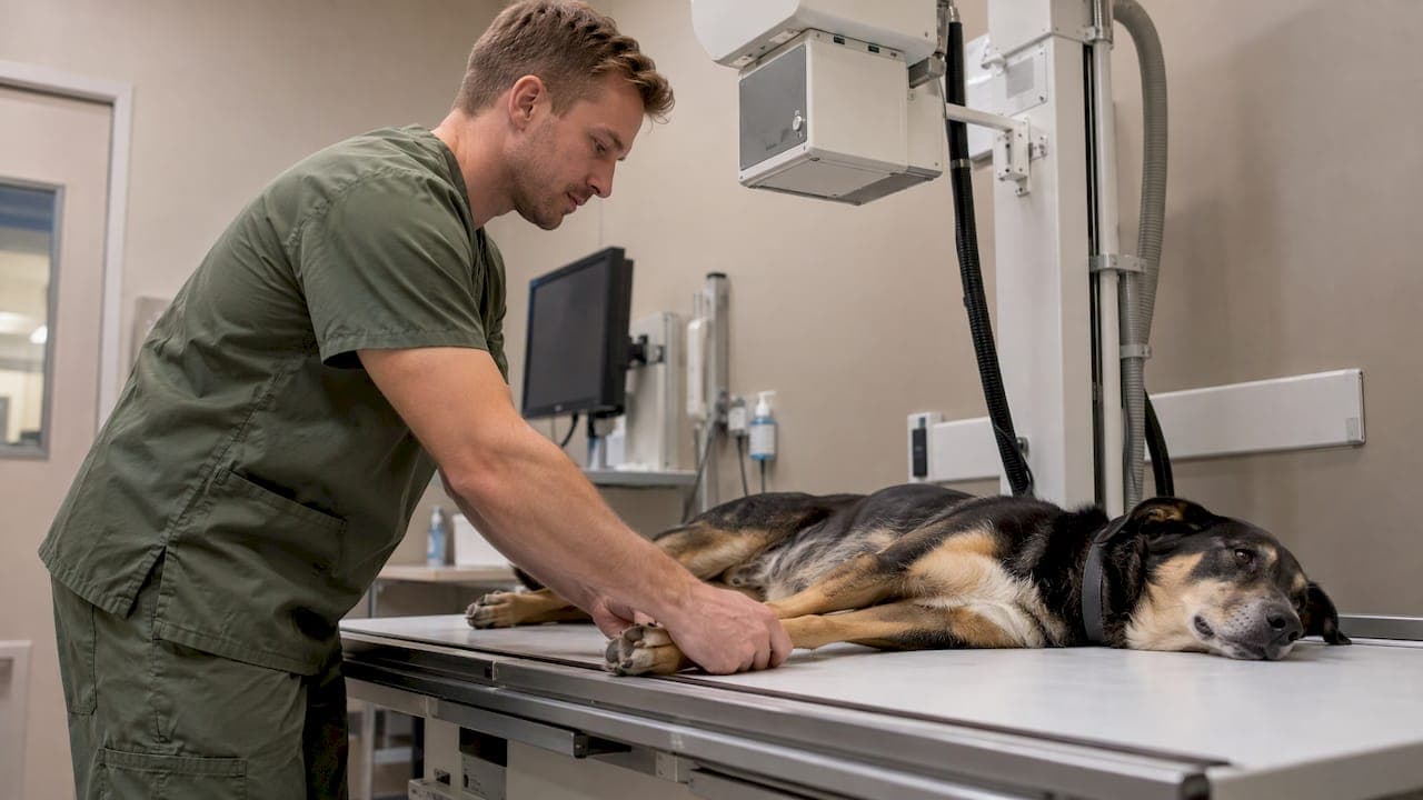
Some dogs lie down for an X-ray awake and calm. Others are sedated so the images come out sharp and the dog stays comfortable. The deciding factors are pain, anxiety and whether your dog can hold still in the position the radiologist needs. Your veterinarian makes that call after examining your dog, and the reasons are usually simple once you see them laid out.
This guide explains when sedation is recommended, when it isn't needed, how sedation differs from general anesthesia, what X-rays and ultrasound can and cannot show, and how to prepare so the visit goes smoothly.
TL;DR
- Many dog X-rays are done awake. Sedation or anesthesia is considered when pain, anxiety or movement would compromise the images.
- Sedation often means sharper pictures, fewer repeat exposures and less manual restraint, which helps your dog and the staff.
- For chest X-rays, light sedation can be a better choice than general anesthesia, because anesthesia can change how the lungs look.
- X-rays show bones and large body cavities well. Ultrasound shows soft tissue and organs in real time. Vets often use both.
- Tell your veterinarian about your dog's fear, pain and medical history before the visit so the plan fits your dog.
Table of Contents
- Do dogs need sedation for an X-ray?
- When a dog can have an X-ray without sedation
- When your veterinarian may recommend sedation
- Sedation versus general anesthesia
- How to prepare your dog
- What a dog X-ray can show
- What an ultrasound shows in dogs
- X-ray vs. ultrasound for dogs
- Imaging at City Animal Veterinary Center
- FAQ
- Sources
Do dogs need sedation for an X-ray?
Not always. Cornell's College of Veterinary Medicine explains that radiographs are often performed awake, sometimes with a sedative, and that sedation or anesthesia is considered when pain, anxiety or movement would defeat the procedure. The 2019 WSAVA congress proceedings add that most chest and abdominal radiographs do not require sedation, though some patients benefit from a sedative or pain medication.
The Merck Veterinary Manual takes a slightly different angle: sedation or short-acting anesthesia is often necessary and usually desirable when medically permitted. The reasons it lists are practical. Sedation reduces anxiety, minimizes manual restraint, cuts down on repeat images and controls pain during positioning.
So the honest answer is that it depends on the dog and on the body part being imaged. A relaxed senior Lab getting a chest film is a different case from a frightened rescue with a sore hip.

When a dog can have an X-ray without sedation
A dog is a good candidate for an awake X-ray when three things line up:
- Calm temperament. A relaxed dog that tolerates gentle handling will usually stay put. A WVS Academy radiography guide notes that a relaxed dog that stays positioned with positioning aids may not need sedation, while an excitable or anxious dog may need sedation or general anesthesia to get diagnostic-quality images.
- Little or no pain. Lying on the back or stretching a limb is easy for a comfortable dog and hard for a dog with a sore joint or injured spine.
- A simple view. Chest and abdomen views are generally easier to get awake than precise orthopedic positioning.
Veterinary teams also use positioning aids such as foam wedges and sandbags so that people are not holding the dog under the beam. Cornell notes that it minimizes manual restraint for radiographs to limit staff radiation exposure. If you want to know how a clinic keeps both pets and people safer during imaging, our guide to pet X-ray safety for owners walks through it.
When your veterinarian may recommend sedation
Sedation tends to come up in a handful of situations.
Pain
The Merck manual points out that painful problems such as fractures and arthritis can make manipulation painful. It also says an acutely paraparetic or paraplegic animal may be hard to position properly for spinal radiographs without anesthesia. Here sedation is as much about comfort as image quality, because pain medication is often part of the plan.
Orthopedic views
Hip, elbow, knee and spine films depend on straight, symmetrical positioning. The 2016 WSAVA orthopedic radiography proceedings recommend sedation highly for orthopedic radiographs because straight positioning and reduced pain and stress improve the examination. If your dog is limping and the vet suggests sedated films, this is usually why.
Anxiety and fear
A dog that is trembling, panting or struggling is hard to image. Movement blurs the picture, which can mean repeating the exposure. PetMD's 2024 guide says sedation is not required for every dog X-ray, but that reducing movement can shorten exposure, prevent blur or distortion, reduce repeat images and limit radiation exposure for the dog and staff.
If vet visits are a struggle for your dog in general, our article on calmer vet visits for anxious dogs gives practical steps to start at home.
Restless or large dogs
Puppies, high-energy adolescents and very large dogs can be tough to position awake. A short period of sedation often makes the whole process quicker and gentler for everyone.
Sedation versus general anesthesia
These are not the same thing. Sedation relaxes your dog and takes the edge off fear, and your dog is still easy to rouse. General anesthesia means full unconsciousness with a protected airway. Most X-rays, if a dog needs help at all, call for sedation rather than anesthesia, and the lighter option is often the better clinical choice.
Chest films show why. A 2023 peer-reviewed review on thoracic radiograph technique says sedation may reduce motion, while general anesthesia can increase atelectasis (partial lung collapse), which can obscure or mimic lung disease. When it is clinically appropriate, sedation may be preferable to general anesthesia for thoracic radiographs. A calm, sedated dog can give a more readable lung image than an anesthetized one.
Older dogs and dogs with heart, kidney or liver conditions need an individual plan. If your dog is a senior, our piece on senior pet anesthesia and what to look for in a clinic covers the questions worth asking. Always tell your veterinarian about every medication and supplement your dog takes, and about any past reaction to sedatives, because the drug choice and dose are matched to the individual dog.

How to prepare your dog
A little planning makes the visit shorter and calmer.
- Follow the clinic's instructions on food and water. If sedation is possible, your veterinarian may give you specific instructions. Follow those exactly rather than guessing.
- Share the history. Mention pain, previous sedation experiences, heart or breathing problems, and any medication your dog takes.
- Bring a calm companion item. A familiar blanket or a favorite treat (if food is allowed) can help.
- Keep the trip itself low-stress. Car rides can wind a dog up before it even arrives. Our guide to calm car rides for dogs offers step-by-step help.
- Plan a quiet recovery. If your dog was sedated, offer a comfortable resting area and limit activity until the effects wear off. Mild soreness can occur when sedation or positioning was involved, and you should call your clinic if your dog seems unusually uncomfortable or does not seem to be recovering as expected.
If getting your dog into a car is the hardest part, a home visit can take the travel out of the equation for the exam and the conversation about next steps.
What a dog X-ray can show
An X-ray is a two-dimensional image of a three-dimensional dog. According to PetMD, dense bone appears whiter, soft organs such as the liver and spleen appear gray, and air appears black. The Merck Veterinary Manual says X-rays show bones, some foreign objects and large body cavities, and can detect fractures, tumors, injuries and deformities.
PetMD lists many reasons a veterinarian may recommend radiographs, including:
- Trauma and fractures
- Tumors or cancer
- Swallowed foreign objects and gastrointestinal obstruction
- Heart and respiratory disease
- Fluid in the chest or abdomen
- Bladder stones and enlarged organs
- Arthritis and hip dysplasia
- Pregnancy and dental disease
- Surgical planning
What an X-ray cannot show well
Standard X-rays do not show soft tissues in fine detail. The Merck manual explains that contrast material given intravenously or by mouth, or air used as a negative contrast, can outline structures such as the kidneys, heart or digestive tract.
Joints are another example. A 2022 review of canine osteoarthritis says radiography remains a standard, accessible method but has limited sensitivity for early disease and cannot reliably show cartilage, synovial fluid or other soft-tissue joint structures on plain films. It also cautions that the severity visible on a film may not match a dog's pain or function, so findings are best read alongside the physical exam.
Because subtle findings can be misread, PetMD recommends review by a board-certified radiologist when needed. A good clinic treats the X-ray as one piece of the picture, never the whole diagnosis.
What an ultrasound shows in dogs
Ultrasound uses sound rather than radiation. The MSD Veterinary Manual explains that a transducer sends short sound pulses through gel-coupled skin, records the strength, return time and direction of the echoes, and turns those data into a real-time image of tissue structures.
Cornell says ultrasound is commonly used to evaluate organ structure, fluid accumulation, blood flow, infection, tumors, trauma and heart function through echocardiography. It is especially useful for soft tissues in the abdomen, musculoskeletal system and chest, though gas and bone reflect or absorb sound and can shadow structures behind them.
Most dogs tolerate ultrasound awake because it is painless and quiet, though the dog must lie still and often needs a shaved patch of fur for a clear view. A calm environment helps a lot, and our Fear Free ultrasound prep guide explains how to get an anxious dog ready.
X-ray vs. ultrasound for dogs
Owners often ask which one is better. Neither wins every time, because they answer different questions.
| X-ray (radiograph) | Ultrasound | |
|---|---|---|
| Best for | Bones, joints (bone changes), some foreign bodies, big-picture view of chest and abdomen | Soft tissue and organs, fluid, blood flow, heart function |
| Image type | Still, two-dimensional | Moving, real time |
| Weaknesses | Poor detail of soft tissue and organ interiors | Gas and bone can block the view |
| Radiation | Brief, low-dose exposure | None |
| Sedation | Sometimes, depending on pain, anxiety and positioning | Often not needed |
The Merck manual sums up the split: ultrasound is better than standard X-rays for soft tissues and gives moving images, while X-rays are better for bones, some foreign bodies and overviews of large cavities. It also says ultrasound is painless, noninvasive and safe, and that X-ray exposure is brief and low-dose, with lead shielding protecting personnel and the rest of the patient's body.
A liver example from the 2015 WSAVA proceedings shows why both are used: survey radiographs can assess liver size and mineralization or gas patterns but not internal architecture, while ultrasound can assess the architecture and neighboring structures such as the portal vein. In practice, a vet may X-ray first for the overview and then use ultrasound to look closer at an organ that looks abnormal.
Our view on sedation and imaging
We see the same pattern again and again: a dog that arrives frightened has a harder time with every part of the visit, and imaging is no exception. A struggling dog means blurry films, repeat exposures and a longer, more stressful day for everyone. That is why we build our visits around Fear Free principles, so we can lower stress before anyone reaches for medication.
When a dog is still too anxious or too sore to hold a position, we believe a carefully chosen sedative, matched to that dog's age, health and temperament, is a kindness rather than a shortcut. It lets us get the information we need once, gently, and get your dog home.
We also like having imaging under one roof. Digital X-rays, ultrasound and in-house lab work let us connect the findings to the exam, so the answer comes from the whole picture, not a single image. Our approach is team-centered: we talk through the plan with you, explain why each step helps your dog, and adjust it if your dog tells us it is too much.
CAVC
Imaging at City Animal Veterinary Center
If your dog needs an X-ray or ultrasound, we offer diagnostic imaging in Brooklyn, including digital X-rays, ultrasound and lab tests, delivered with our Fear Free approach to keep visits as stress-free as possible for you and your dog.

If your dog is limping, has swallowed something it shouldn't have or seems unwell right now, our urgent care team can help. If travel is hard for you or your pet, ask about our home visits. To start with a conversation about your dog's needs, visit City Animal Veterinary Center and book an appointment.
FAQ
Are dogs sedated for X-rays?
Some are and many are not. Sedation is considered when pain, anxiety or movement would make it hard to get clear images, and it is common for orthopedic films of the hips, elbows and spine. Calm dogs having chest or abdominal X-rays are often imaged awake with positioning aids.
Can a dog have an X-ray without sedation?
Yes, if your dog is calm, comfortable and able to lie in position. Veterinary references note that many radiographs are done awake. The trade-off is that a dog who moves may need repeat images, so your veterinarian may suggest a sedative to get it right the first time.
Does a dog X-ray require anesthesia?
Usually not. Most dogs who need help staying still receive sedation, which is lighter than general anesthesia. For chest X-rays in particular, sedation can be preferable because general anesthesia may cause lung changes that look like disease on the image.
What do dog X-rays show?
They show bones, some foreign objects and the size and shape of organs within large body cavities, so they help find fractures, tumors, deformities and some obstructions. They do not show the fine internal detail of soft tissue organs, which is where ultrasound or contrast studies come in.
What does a dog ultrasound show?
It shows organ structure, fluid, blood flow and heart function in real time. It is the go-to tool for looking inside the liver, kidneys, bladder, spleen and other soft tissues. Gas and bone can block the view, so it complements X-rays rather than replacing them.
Sources
- Imaging Service, Cornell University College of Veterinary Medicine
- Radiography of Animals, Merck Veterinary Manual
- Diagnostic Imaging, Merck Veterinary Manual (Special Pet Topics)
- Ultrasonography in Animals, MSD Veterinary Manual
- Radiographic Signs of Common Bone and Joint Disease in Dogs, WSAVA 2016
- Techniques for Chest and Abdomen: Awake and Sedate, WSAVA 2019
- Thoracic Radiograph Technique, Today's Veterinary Practice, 2023
- X-Rays for Dogs, PetMD
- Preparing the patient, WVS Academy
- Moving Beyond the Limits of Detection: canine osteoarthritis imaging, PMC, 2022
- The Liver: One Big Brown Organ in Gray Shades, WSAVA 2015