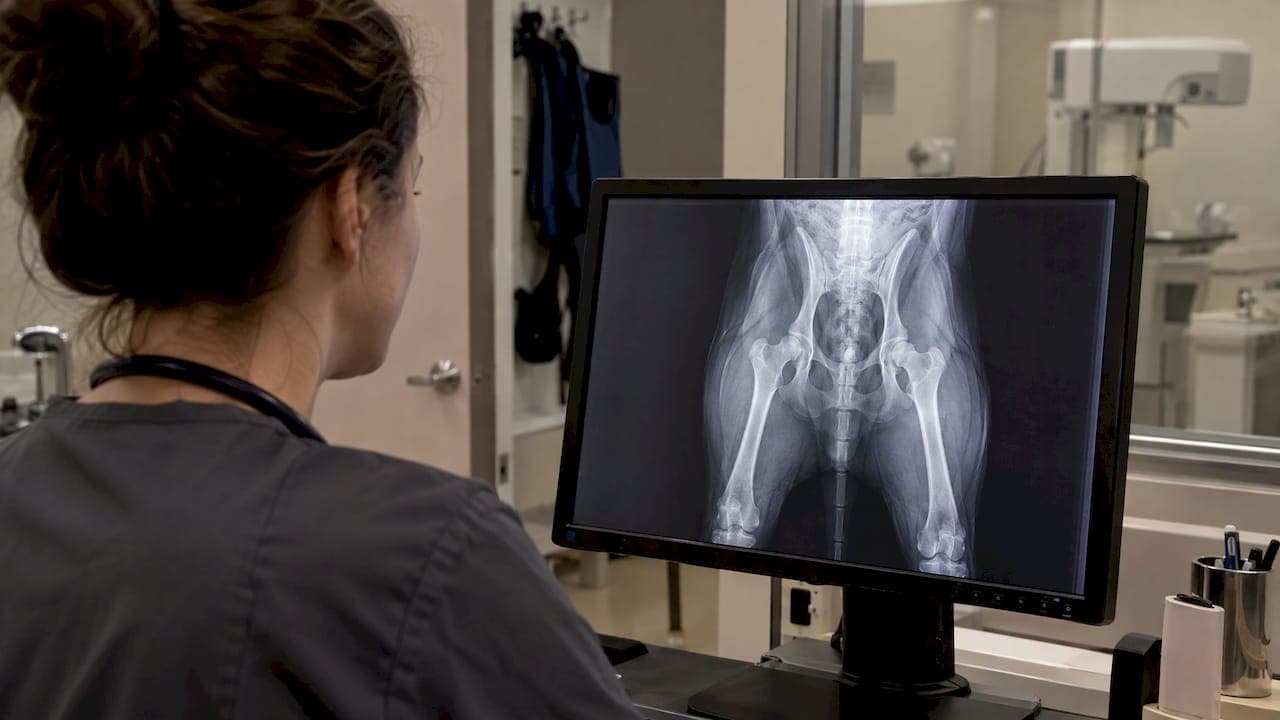
A hip X-ray lets your veterinarian see how the ball of your dog's thigh bone sits in the socket of the pelvis, and whether the joint has already started to change shape. It is the central test for diagnosing canine hip dysplasia, but it works best alongside a hands-on exam, and the picture only makes sense when the dog is positioned correctly. This guide explains what a vet looks for on the film, how the images are taken, how to read a hip score, and what to ask once you have a result.
TL;DR
- Hip dysplasia is diagnosed with radiographs plus a physical exam and hip palpation, and the positioning for the images generally requires sedation or anesthesia, according to Cornell's College of Veterinary Medicine.
- On the image, vets look at how well the femoral head sits in the socket, the shape of both, and signs of arthritis.
- OFA grades hips in seven categories, and PennHIP measures hip laxity as a number. They answer different questions.
- An X-ray finding does not by itself prove that your dog's limp comes from the hips, so the whole dog gets examined.
- Bring your questions about activity, comfort and next steps to the appointment. We cover the main ones below.
Table of Contents
- What a hip X-ray shows in a dog
- How vets diagnose hip dysplasia: exam first, then images
- How the hips are positioned
- OFA scores explained
- PennHIP: measuring looseness in young dogs
- What an X-ray score can and cannot tell you
- After the diagnosis: questions to bring to your vet
- How to prepare your dog for a hip X-ray
- Care for your dog's hips at City Animal Veterinary Center
- FAQ
- Sources
What a hip X-ray shows in a dog
In a healthy hip, the round head of the femur sits deeply and snugly in the acetabulum, the cup-shaped socket in the pelvis. In hip dysplasia the fit is loose, so the joint wears abnormally over time.
The World Small Animal Veterinary Association (WSAVA) radiology guidance describes the earliest sign as a femoral head that is not well seated in the socket, followed by abnormal wear, bone remodeling and degenerative joint disease. As the disease progresses, the same source lists these findings on radiographs:
- Partial or complete displacement of the femoral head (subluxation or luxation)
- Loss of the round shape of the femoral head
- A shallow or remodeled socket
- An irregular socket edge
- New bone growth around the joint (periarticular osteophytes)
- Morgan's line, a bony thickening along the neck of the femur
- Subchondral sclerosis, a denser look to the bone under the joint surface
DVM360 conference proceedings explain that the earliest abnormality on an extended hip view is subluxation, defined there as less than 50% of the femoral head covered by the dorsal rim of the socket. The same source notes that persistent instability can produce Morgan's line, while progressive degenerative disease may flatten the femoral head, make the socket shallower and add osteophytes.
In plain terms: early on, the X-ray shows a loose fit. Later, it shows the bone responding to years of that looseness.
Why vets also use X-rays to rule things out
Radiographs do more than confirm dysplasia. DVM360 notes they objectively evaluate conformation and degenerative change, and with specialized techniques, laxity. They also help rule out other causes of hind-limb lameness. A limp that looks like a hip problem can come from the knee, the spine or soft tissue, so the images answer two questions at once: what is going on in the hip, and what else could explain the signs.
If your dog is limping and you are not sure why, our guide to limping dog causes explains what is behind it and when to call a vet.
How vets diagnose hip dysplasia: exam first, then images
The X-ray is one part of a larger workup. Cornell states that canine hip dysplasia is diagnosed with radiographs plus a physical examination and hip palpation, including the Ortolani sign.
The physical exam
Your veterinarian will watch your dog stand and walk, feel the muscles of the hindquarters, and move each hip through its range of motion. Signs owners often notice at home include trouble rising, reluctance to climb stairs, a swaying or "bunny hopping" gait, and stiffness after rest.
The Ortolani test checks for a loose hip. As DVM360 describes it, the examiner pushes along the femur to subluxate a lax hip, then abducts the leg until the femoral head slips back into the socket. Sedation improves confidence in the result, because muscle tone in an awake dog can mask the finding.
Why sedation is part of the picture
Good hip films need a relaxed dog. A tense dog cannot be placed symmetrically, and a hip that hurts is hard to extend flat. Cornell notes that the special positioning generally requires sedation or anesthesia. For an anxious dog, a calm, low-stress setting makes this far easier on everyone. Our piece on pet X-ray safety for owners explains how images can be taken safely without owners holding their pets.
How the hips are positioned
Positioning is not a detail. It determines what the image can tell you.
For the standard view used by the Orthopedic Foundation for Animals (OFA), the dog lies on its back with the rear legs extended and parallel, the stifles (knees) rotated inward and the pelvis symmetric. OFA recommends chemical restraint to the point of relaxation.

This extended view is the long-standing standard, but it has a known quirk. DVM360 points out that extension and inward rotation tighten the joint capsule, so the view can understate laxity. A normal-looking extended image therefore does not exclude clinically relevant laxity. That is one reason your vet pairs the picture with the exam, and why some practices use additional techniques for young dogs.
What your dog needs from the process
- A quiet room and calm handling, since stress makes muscles tighten
- Sedation or anesthesia chosen by your veterinarian for your dog's age, health and temperament
- Correct, symmetric positioning, repeated if the first image is crooked
- Enough time for the dog to recover afterward in a comfortable place
This is where a Fear Free approach matters. Dogs that associate the clinic with fear tend to resist handling, and resistance makes positioning harder. Pre-visit calming steps, gentle restraint and treats are practical tools, not extras.
OFA scores explained
OFA is a registry that grades hip radiographs. It grades hips in seven categories: Excellent, Good, Fair, Borderline, Mild, Moderate and Severe, and the final grade reflects consensus among three independent radiologists.
| OFA grade | What it means |
|---|---|
| Excellent | Within normal limits; superior hip conformation |
| Good | Within normal limits; a well-formed, congruent joint |
| Fair | Within normal limits; minor irregularity, with slightly increased joint width or coverage changes |
| Borderline | Not clearly normal or dysplastic |
| Mild, Moderate, Severe | Dysplastic, with increasing degrees of change |
The first three rows come directly from OFA's grade definitions. A "good hip score" in everyday language means one of those three.
When can a dog be scored?
Age matters. Purdue's Canine Welfare Science resource states that OFA screening may be preliminary from 12 months and definitive from 24 months, while PennHIP can assess dogs from 4 months of age.

PennHIP: measuring looseness in young dogs
PennHIP takes a different approach. Instead of grading the shape of the joint, it measures how far the femoral head can be pulled out of the socket. Cornell identifies the PennHIP distraction method as the most accurate measurement of puppy hip laxity and says participating veterinarians and technicians must be certified.
The American College of Veterinary Surgeons describes it as a quantitative measurement of actual hip laxity and the most accurate early-age radiographic method, and notes that specialized training and equipment are required. It can help predict future arthritis and inform surgical options, whereas standard OFA screening is less predictive at very young ages.
The American Kennel Club's veterinary-authored guide puts the contrast simply: OFA gives a qualitative assessment, while PennHIP gives a number estimating the risk of painful osteoarthritis later in life.
Which one fits your dog?
| Question | OFA | PennHIP |
|---|---|---|
| What does it assess? | Joint shape and arthritic change | Laxity, as a measured number |
| Youngest reliable age | Preliminary at 12 months, definitive at 24 months | 4 months |
| Who performs it? | Any veterinarian can take the films for submission | Certified veterinarians and technicians |
| Typical use | Screening and diagnosis in adult dogs | Early risk assessment in puppies |
If you have a puppy of a large breed and want an early read, ask your veterinarian whether early-age assessment makes sense or whether a referral to a certified provider is the better route. If you have an adult dog with symptoms, standard radiographs read by your veterinarian are usually the starting point.
What an X-ray score can and cannot tell you
A hip score is a snapshot of the joint. It is useful, and it is also not a crystal ball.
It does not match symptoms perfectly. The AKC guide notes that hip films give insight into the hip phenotype, but no available test perfectly predicts which dogs will develop clinical signs of arthritis. Some dogs with significant changes on film move comfortably for years, and others with milder changes are sore.
It does not prove a limp is from the hips. Veterinary Partner advises that dysplasia and arthritis on an X-ray do not by themselves prove a dog's hind-end pain or lameness comes from the hips, so other causes should be evaluated. A sudden new limp deserves a fresh exam, not an assumption.
It is progressive once arthritis appears. OFA's framework states that once secondary arthritis is reported, it continues to progress over time. That is a reason to build a long-term comfort plan early rather than wait.
It matters for breeding, with limits. Scores help reduce inherited risk, but the AKC's author emphasizes that even dogs with normal hips can produce affected puppies.
After the diagnosis: questions to bring to your vet
A diagnosis is the start of a plan. These questions help you get a useful one:
- How severe are the changes, and what is my dog's grade or measurement? Ask to see the images and have the findings pointed out.
- Are the hips the main source of the discomfort? Given the point above, this is worth asking directly, especially if your dog also has knee or spine concerns.
- What is the plan for comfort? Options your veterinarian may discuss include weight management, controlled exercise, joint-support measures, pain medication, and complementary therapies.
- Is surgery something to consider now or later? Your vet or a board-certified surgeon can explain which procedures suit a dog's age and joint condition.
- How often should we recheck? Regular exams catch changes in comfort before they become big setbacks.
- What can I change at home? Non-slip flooring, ramps and a supportive bed make daily life easier.
Veterinary Partner notes that owners should discuss routine screening based on the dog's intended use and concerns, and that X-rays are appropriate when hind-end lameness or hip-pain signs are present.
Supporting an arthritic hip at home
If your dog is already showing arthritis, daily comfort measures add up. Short, steady walks are usually kinder to a hip than occasional hard play. Keep your dog lean, since extra weight loads every joint. Provide traction on slick floors. For some dogs, acupuncture is part of the comfort plan, and we have written about acupuncture for arthritic dogs in Brooklyn, including how it works at home visits. Our pain management service covers the medical side of keeping a dog comfortable.
Senior dogs often carry the hip changes alongside other age-related issues, so a thorough senior dog wellness exam is a good time to review mobility.
How to prepare your dog for a hip X-ray
A little preparation makes the visit smoother.
- Follow the fasting instructions your clinic gives you. If sedation or anesthesia is planned, you will be told when to stop food and water.
- Tell us about medications and supplements. Include anything your dog takes, since some drugs affect sedation choices.
- Mention any past reactions. If your dog has had trouble with sedation before, say so.
- Bring a favorite blanket or treat. Familiar comforts lower stress.
- Plan a quiet recovery. Your dog may be groggy for the rest of the day, so keep the evening calm.
Image quality has improved with digital systems. If you are curious about the technology, our article on digital X-ray versus film for pets explains why careful technique and dose still matter.
Our perspective on hip X-rays
We see hip X-rays as one of the most useful conversations a vet can have with an owner, because the image gives you something concrete to look at together. What we care about just as much is how the dog experiences getting there.
A dog that is frightened will tense up, and a tense dog is hard to position well. That is why we build our visits around Fear Free principles: calm handling, patience and a setting that lowers stress for pets and owners alike. A good image starts with a settled dog.
We also believe the picture should never stand alone. We pair radiographs with a careful physical exam, and we look at the whole dog, because comfort is what you are really trying to protect. For dogs with arthritic hips, we bring our holistic therapy options, such as acupuncture and pain management, alongside traditional veterinary care, so the plan fits your dog instead of just the film.
For owners who find the trip hard, our home visit service is there so your dog's care does not stop at the front door.
CAVC
Care for your dog's hips at City Animal Veterinary Center
At our Brooklyn practice, we offer diagnostic imaging including digital X-rays and ultrasound, an in-house laboratory, pain management, acupuncture and surgery, all delivered with a Fear Free approach designed to keep visits calmer for anxious pets. If your dog is slowing down, struggling with stairs or limping, start with a wellness exam so we can examine your dog and talk through whether hip imaging is the right next step.

If getting your dog to the clinic is the hard part, ask about our home visit and house call service. If your dog has sudden, severe pain or cannot bear weight, our urgent care team can see you.
FAQ
What does a hip dysplasia X-ray show?
It shows how well the femoral head fits in the socket and whether the joint has changed shape. Findings can include subluxation, a flattened femoral head, a shallow socket, new bone growth around the joint and Morgan's line. The same images help rule out other causes of hind-limb lameness.
Does my dog need to be sedated for a hip X-ray?
Usually, yes. Cornell states that the special positioning generally requires sedation or anesthesia. Relaxation lets the vet extend and align the legs correctly, and it also makes the hip exam more reliable because muscle tone can hide looseness in an awake dog. Your veterinarian will choose the approach that fits your dog's age and health.
What is a good hip dysplasia score?
On the OFA scale, Excellent, Good and Fair are all within normal limits. Borderline sits between normal and dysplastic, and Mild, Moderate and Severe indicate dysplasia. PennHIP works differently: it yields a measured laxity number that helps estimate later arthritis risk.
Can an X-ray tell me how my dog will do in the future?
It gives useful clues, but not a guarantee. The AKC's veterinary guide notes that no available test perfectly predicts which dogs will develop clinical arthritis signs. Your vet combines the image with your dog's exam, age, weight and activity to build a realistic outlook.
At what age can hip dysplasia be seen on X-ray?
Purdue's Canine Welfare Science resource states that PennHIP can assess dogs from 4 months. OFA screening may be preliminary from 12 months and definitive from 24 months. Dogs of any age with limping or hip pain can be X-rayed to look for changes.
Sources
- Hip Dysplasia | OFA
- Hip Grade Details | OFA
- Canine hip dysplasia (CHD) | Cornell University College of Veterinary Medicine
- Radiological Signs of Hip Dysplasia | WSAVA 2016 Congress
- Diagnosing hip dysplasia (Proceedings) | DVM360
- Canine Hip Dysplasia | Purdue Canine Welfare Science
- Canine Hip Dysplasia | American College of Veterinary Surgeons
- Genetics of Hip Dysplasia in Dogs: A Guide for Breeders | AKC
- Hip Dysplasia in Dogs: Background, Signs and Diagnosis | Veterinary Partner