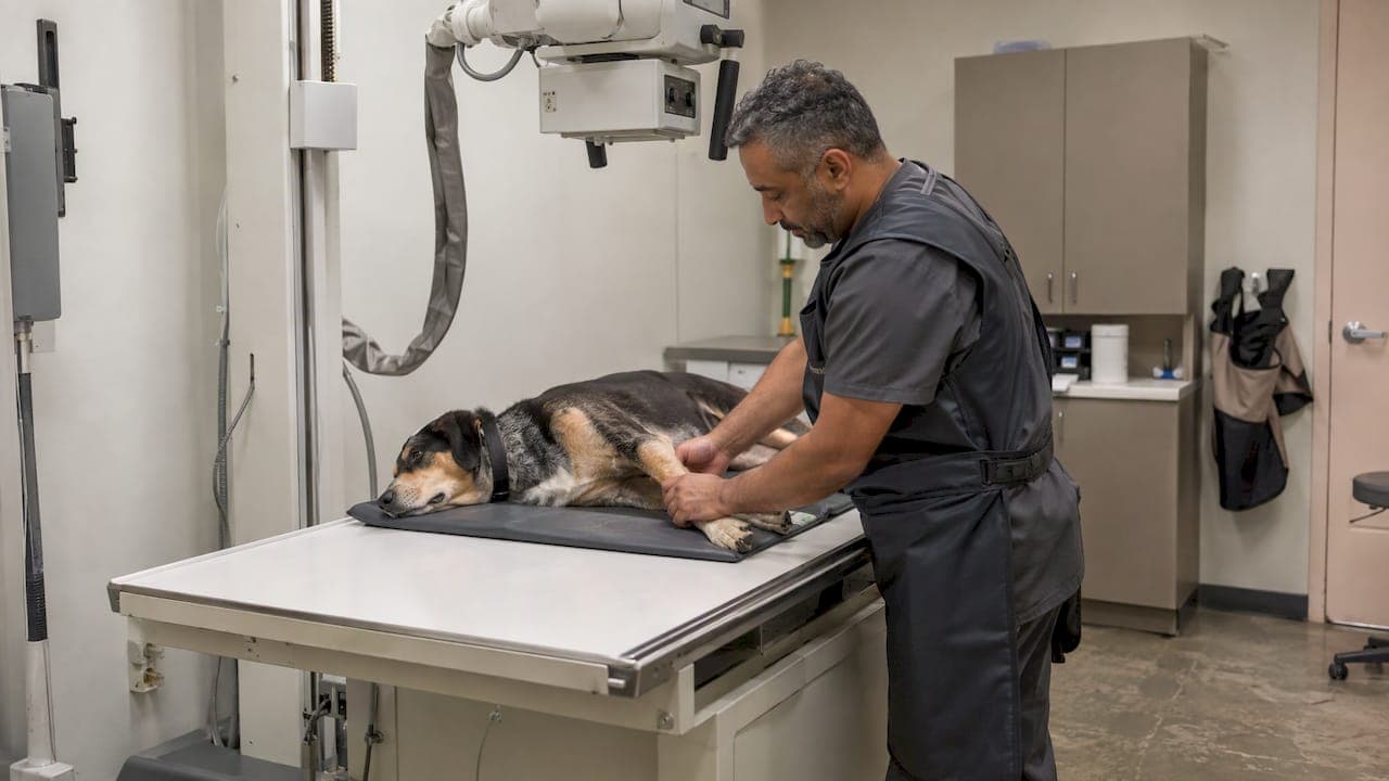
Orthopedic X-rays (radiographs) are pictures of your dog's bones and joints. Vets use them to find the cause of limping, to check hips, knees, elbows and legs after an injury, and to plan surgery. The study is painless and usually takes only minutes. The main thing you can do is help your dog stay calm, because good positioning is what makes the images useful. If you've been wondering about a dog leg X-ray, this guide covers why it's recommended, what it can show, how to prepare and what comes next.
TL;DR
- Vets recommend orthopedic X-rays to find the cause of lameness, check joints and growth-plate problems, assess hips and knees, and plan surgery.
- A leg X-ray shows bone and joint changes well. Ligaments and tendons are harder to judge, so your vet may add an exam, ultrasound or other imaging.
- Sedation is often used so your dog can be positioned straight and comfortably. That often means fewer repeat images.
- Ask whether to withhold food beforehand if sedation may be needed, and follow any medication instructions exactly.
- Our Fear Free approach is built to keep anxious dogs calmer, from the exam room to the X-ray table.
Table of Contents
- Why a vet recommends orthopedic X-rays
- What a dog leg X-ray can show
- When the X-ray is only the first step
- Why dogs are sometimes sedated
- How the X-ray session goes
- How to prepare your dog
- Are X-rays safe for dogs?
- What happens after the images
- Perspective: why calm matters for good images
- Orthopedic care at City Animal Veterinary Center
- FAQ
- Sources
Why a vet recommends orthopedic X-rays
Lameness has many possible causes, and a physical exam alone can't always separate them. During an orthopedic exam, a vet looks for swelling, fluid in a joint, pain, instability, a grating sensation called crepitus, reduced range of motion and muscle loss. The MSD Veterinary Manual lists these findings and notes that imaging can help diagnose the cause of lameness.
The usual reasons for ordering images:
- A dog that limps and won't improve. If you're trying to work out what's behind it, our guide to limping dog causes explains when to call a vet.
- Trauma. After a fall, a car accident or a bad landing, X-rays check for fractures and dislocations. The American College of Veterinary Surgeons (ACVS) explains that hip radiographs can show whether a hip is dislocated, in which direction, and whether the joint surface is fractured. Extra views may be taken to look at the chest, spine, abdomen or other limbs.
- A growing puppy with a lame leg. Veterinary Partner says radiographs should be taken in a lame growing dog to screen for conditions such as osteochondritis dissecans, elbow dysplasia, patellar luxation and growth-plate fractures.
- Hip screening. ACVS says specially positioned, lightly sedated X-rays help diagnose hip dysplasia and measure hip laxity. Early results can help decide whether preventive surgery makes sense.
- Suspected arthritis. Stiffness after rest, trouble with stairs and reluctance to jump all prompt a look at the joints.
- Before surgery. Images help the surgeon plan and, when needed, measure for implants.
What a dog leg X-ray can show
Radiographs are at their best with bone. They show the outline, density and alignment of the bones and the spacing of the joints.
| Problem | What the X-ray may reveal |
|---|---|
| Fracture or dislocation | Breaks, displaced bones, a joint out of position |
| Osteoarthritis | Bone spurs, joint swelling, denser bone beneath the cartilage, mineral within the joint, bone cysts |
| Developmental disorders | Angular deformity, prematurely closed growth plates |
| Hip dysplasia | Shape of the hip and how loose the joint is |
| Knee problems | Swelling in the joint, kneecap position, bone shape in the rear leg |
For arthritis, ACVS reports that radiographs may show osteophytes, joint effusion, soft-tissue swelling and subchondral sclerosis, among other signs. For growth problems, the MSD Veterinary Manual describes radiography as useful for developmental bone disorders, including angular deformities and early growth-plate closure.
Why vets take more than one view
A single picture is flat, so a bone can hide a problem behind itself. The MSD Veterinary Manual recommends orthogonal views of the affected bone or joint, meaning two images at right angles, sometimes compared with the opposite limb. If your vet X-rays both legs when only one is limping, that is the reason. Your dog's healthy side gives a baseline for what normal looks like for that dog.

Pictures plus the exam
What you see on an image has to match what the vet feels and observes. Zoetis stresses that arthritis assessment is more reliable when orthogonal views, side-to-side comparison and clinical examination are considered together. That is why we pair imaging with a hands-on orthopedic exam rather than treating the picture as the whole story.
When the X-ray is only the first step
Bones are the X-ray's strength. Ligaments, tendons and cartilage are soft tissue, which radiographs show less clearly. PetMD notes that X-rays can't definitively diagnose cruciate ligament or meniscal damage. For a limping dog with a suspected knee injury, they still do real work: they check the joint for swelling and arthritis, rule out other causes and help plan any surgery.
If the first images don't explain the problem, the next step is usually another kind of imaging:
- Ultrasound shows tendons and muscle directly. For Achilles problems, ACVS says X-rays evaluate the ankle joint and heel bone, while ultrasound visualizes the tendon itself.
- CT gives high-contrast detail of bone. The MSD Veterinary Manual says CT and MRI can add bone and soft-tissue detail that plain films can't.
- MRI helps show soft-tissue and joint injuries.
We offer digital X-rays and ultrasound on site, along with an in-house laboratory, so many first steps can happen in one visit. If your vet suggests ultrasound, our ultrasound prep guide walks through what to expect. If a case needs advanced imaging such as CT or MRI, we will explain why and what it adds.
Why dogs are sometimes sedated
A dog in pain doesn't want its leg stretched out on a hard table. A dog that's anxious will squirm. Both make a crooked, blurry image, and a crooked image can mean a repeat.
VIN says sedation can make positioning less stressful and painful, allow more thorough palpation and manipulation, and support radiation safety. ACVS notes that for rear-leg studies of the pelvis, knee and tibia, sedation is often needed for straight views and comfort. The MSD Veterinary Manual adds that fractious patients may need sedation for palpation and radiography.
Sedation isn't automatic. A calm, cooperative dog with a simple question may need nothing. A dog being screened for hip dysplasia or one with a painful limb often does. Your vet decides, based on your dog's age, health and temperament. If your dog has a heart condition, kidney disease or is a senior, tell us before the visit. Our guide to senior pet anesthesia covers what good safety practice looks like.
How the X-ray session goes
- Exam first. The vet watches your dog walk, feels the limbs and joints, and decides which areas to image.
- Positioning. According to PetMD, dogs may be placed on their side, back or belly and supported with troughs, sandbags, foam wedges or tape.
- Calibration, when needed. For surgical planning, VIN explains that a radiopaque object of known size placed beside the target lets the team correct for magnification and take accurate measurements.
- Exposure. The exposure itself takes about a second, and PetMD says the overall procedure generally takes only a few minutes.
- Review. The vet reads the images, often alongside your dog's exam findings and any lab results, and talks through options.
Most of the clock time goes into settling your dog and getting the position right, not into the picture itself.
How to prepare your dog
Follow fasting instructions. PetMD says owners may be asked not to feed their dog for several hours before a scheduled study if sedation could be needed. Ask us when you book. Don't guess, and give medications only as your vet directs.
Bring the history. Note when the limp started, whether it's worse after rest or exercise, which leg it is, and any past injuries. A short phone video of your dog walking or trotting at home can show a limp that disappears in a busy exam room.
Bring records. Past X-rays, vaccination records and a list of medications and supplements help us compare and choose safe sedation.
Plan for a calmer visit. Bring a favorite blanket or treats, and let us know how your dog reacts to handling. Our tips on calmer vet visits for dogs are worth reading ahead of time.

Don't force exercise. If your dog is limping, keep activity gentle until you've been seen. Don't give human pain medication, which can be dangerous for dogs. Call us first.
Are X-rays safe for dogs?
Radiographs are painless and noninvasive. PetMD describes them as low-risk overall, while noting that ionizing radiation can damage cells and that limiting movement reduces repeat exposures. That's why positioning and sedation matter.
The American College of Veterinary Radiology says exposure should follow ALARA, As Low As Reasonably Achievable, while preserving diagnostic quality. The same statement says people not essential to the procedure should leave the room, and that staff who must stay should wear protective gear and use dosimetry.
This is also why many practices ask owners to wait outside during the exposure. You can read more in our posts on pet X-ray safety for owners and digital X-ray versus film.
What happens after the images
The outcome usually falls into one of a few paths:
- A clear diagnosis. A fracture, dislocation or advanced arthritis may be visible straight away, and the vet can discuss treatment.
- A partial answer. The images may rule out some causes and point toward soft tissue. The next step may be ultrasound, further tests or a recheck.
- Surgical planning. If surgery is likely, the images feed into the plan. See our surgery services.
- Ongoing joint care. For arthritis, treatment may involve pain management, weight and nutrition guidance and, at our practice, holistic options such as acupuncture alongside conventional care.
One caution about reading results yourself: an image can look worse or better than your dog feels. In arthritis, bone changes and symptoms don't always line up. Treat the X-ray as one piece of information, and let your vet combine it with the exam.
Perspective: why calm matters for good images
We see it every week: the quality of an orthopedic X-ray depends on how relaxed the dog is. A scared dog tenses, pulls away and fights the positioning. A calm dog lets us get the clean, straight views that answer the question the first time.
That's the reason we built our practice around Fear Free handling. Slow introductions, gentle restraint, treats and, when it's appropriate, light sedation all serve the same goal: a better image with less stress for your dog and for you.
We also like to keep the whole workup under one roof. Digital X-rays, ultrasound and in-house lab work let us move from a limp to answers without sending you across the borough. For dogs that can't easily travel, such as seniors or those with severe anxiety, we also offer home visits for the exam portion of care.
CAVC
Orthopedic care at City Animal Veterinary Center
If your dog is limping, favoring a leg or slowing down on walks, we can examine them and take X-rays at our Brooklyn practice. Our Fear Free approach is designed to reduce stress at every step, and our team can talk through sedation, positioning and next steps before anything is done.

Start with a wellness exam to have your dog's joints checked. If the limp is sudden or your dog is in obvious pain, contact our urgent care team. If getting to us is hard, ask about home visits. Many other services, including diagnostic imaging, pain management and geriatric care, are listed on our services page.
FAQ
Does my dog need to be sedated for an orthopedic X-ray?
Not always. Calm dogs with simple questions may be imaged awake. Sedation is common when a limb is painful or when exact positioning is needed, such as for hip screening. VIN notes it can make positioning less stressful and support radiation safety. Your vet will decide based on your dog's health and temperament.
How long does a dog leg X-ray take?
The exposure itself takes about a second, and PetMD says the whole procedure generally takes only a few minutes. Settling your dog and positioning the leg take most of that time. If sedation is used, plan for extra time for your dog to wake up fully.
Can an X-ray show a torn ligament in my dog's knee?
Not directly. X-rays show bones and joint changes well, but PetMD notes they can't definitively diagnose cruciate ligament or meniscal damage. Your vet combines the exam with the images, and may add other imaging if needed.
Why would my vet X-ray the leg that isn't limping?
Comparing both sides gives a baseline for your dog's normal anatomy. The MSD Veterinary Manual says orthogonal views of the affected bone or joint are recommended, sometimes with comparison to the opposite limb. It helps separate a true abnormality from your dog's natural variation.
Can I stay with my dog during the X-ray?
Usually not during the exposure. The American College of Veterinary Radiology says people not essential to the procedure should leave the room. You can stay with your dog for the calming part beforehand, and we will bring them back to you as soon as they are ready.
Sources
- Orthopedic Examination in Small Animals, MSD Veterinary Manual
- Hip Luxation, American College of Veterinary Surgeons
- Canine Hip Dysplasia, American College of Veterinary Surgeons
- Osteoarthritis in Dogs, American College of Veterinary Surgeons
- Patellar Luxation in Dogs, American College of Veterinary Surgeons
- Achilles Tendon Injuries, American College of Veterinary Surgeons
- Developmental Osteopathies in Dogs and Cats, MSD Veterinary Manual
- Osteochondritis Dissecans in Dogs, Veterinary Partner
- Avoiding the Dogogram: Orthopedic Radiography 101, VIN
- X-Rays for Dogs, PetMD
- Tips to Identify and Interpret Canine Radiographs, Zoetis
- ACVR Radiation Safety Statement, American College of Veterinary Radiology