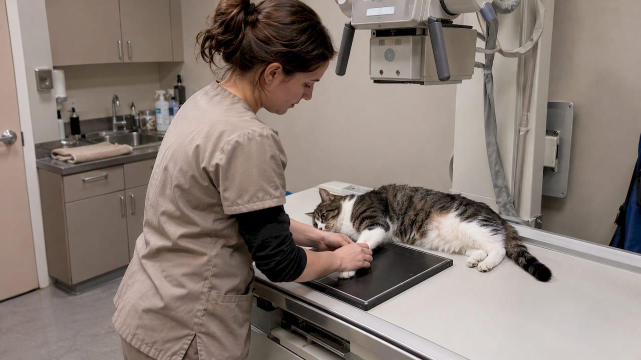
A digital X-ray lets your veterinarian see your pet's bones, chest, abdomen and, with dental films, the roots of the teeth, and view the image on a screen within moments. It is painless, and it is usually one of the first tools a vet reaches for when a pet limps, coughs, vomits or needs a dental check. This guide explains what digital X-rays can and cannot show, how safety is handled, when a pet needs sedation, and what the visit looks like from start to finish.
TL;DR
- Digital X-rays capture an image with an electronic detector instead of film, so the image appears on a screen and is easy to store and share with specialists.
- They are best at showing bones, the chest, many foreign objects and the size and position of organs. Soft-tissue detail sometimes calls for ultrasound or advanced imaging.
- Dental X-rays show what lies below the gumline, where most of a tooth sits, and are normally taken under general anesthesia.
- Most body X-rays need no preparation. Calm handling, and sometimes sedation, is what gets a clear image.
- Radiation safety rests on the ALARA principle: keep exposure as low as reasonably achievable through time, distance and shielding.
Table of Contents
- What a digital X-ray is and how it works
- What X-rays can help your vet find
- Dental X-rays: seeing below the gumline
- What to expect: before, during and after
- When an X-ray is only the first step
- When to call the vet
- Digital X-rays at City Animal Veterinary Center
- FAQ
- Sources
What a digital X-ray is and how it works
An X-ray (radiograph) passes a small, controlled beam through the body. Dense tissue such as bone absorbs more of the beam and shows up white. Air-filled tissue such as lung shows up dark. Soft tissues land in between.
The difference between digital and film is how the image is captured. In a digital system, a detector, usually a flat panel placed behind the patient, converts the X-rays that pass through the animal into an electrical signal that becomes the picture. There is no film to develop.
The Merck Veterinary Manual's guidance on radiography notes that digital radiography is now standard, and that most digital systems installed today are direct radiography (DR) systems. DR is faster than computed radiography (CR) and needs no intermediate reading step.
What that means for you:
- Speed. The image is on the screen right away, so the vet can tell whether the pet was positioned well and retake a view if needed.
- Sharing. Modern veterinary X-rays are usually produced and stored digitally, which makes them easy to share with specialists for a second opinion.
- Records. Images sit with your pet's chart, so a future visit can be compared against earlier films, which matters for slow-changing problems such as arthritis or a heart that is gradually enlarging.
If you are curious about the finer technical points, including why careful technique still matters with digital systems, we cover them in Digital X Ray vs Film for Pets: Why Dose Creep Still Matters.
What X-rays can help your vet find
Radiographs are a screening and staging tool. They answer questions about structure: is something broken, enlarged, displaced, blocked or out of place?
Bones and joints
The Merck Veterinary Manual says radiographs are useful for showing bones, some foreign objects and large body cavities, and that they can detect fractures, tumors, injuries and deformities. For a dog that limps or a cat that suddenly stops jumping, X-rays show fractures, joint changes and growth abnormalities. Our articles on orthopedic X-rays for dogs and hip dysplasia X-rays go deeper on what those images show and how to prepare.
Heart and lungs
Chest films are a standard part of a cough or breathing work-up. The Merck Veterinary Manual reports that chest X-rays frequently help diagnose heart disease in dogs by showing generalized or chamber-specific heart enlargement and fluid in the lungs, and they can be used to monitor congestive heart failure.
Stomach and intestines
When a pet vomits repeatedly or may have swallowed something, abdominal films come first. Today's Veterinary Practice describes abdominal radiography as a first-line screening test for mechanical intestinal obstruction in dogs and cats, with the vet looking for signs such as dilated bowel, stacked loops and abnormal contents.
Cancer work-ups
Cornell's College of Veterinary Medicine notes that survey radiographs can detect cancer metastasis and show possible bone involvement, and can help locate oral or nasal masses. In other words, X-rays often help answer how far a problem has spread, not just whether it exists.
Dental X-rays: seeing below the gumline
If you searched for dental X-rays for pets, this is the part that matters most. When you look into your pet's mouth you see only the crown of each tooth. The FVMA states that most of a tooth's structure lies below the gumline and cannot be assessed visually, which is why it recommends full-mouth radiographs for every dental patient.
Dental films can reveal:
- Infection or abscesses at the tooth root
- Bone loss around a tooth
- Fractured teeth and retained roots
- Resorptive lesions, which are especially common to watch for in cats
A tooth can look nearly normal on top and be badly diseased underneath. That is why a cleaning alone, without images, can leave real problems undiscovered. Our dental team uses radiographs as part of a thorough dental visit; you can read about our dental care and, for cat owners, how to spot tooth resorption early and get Fear Free radiographs. For a step-by-step view of a cleaning, see what a professional cat dental cleaning involves.
Why dental X-rays need anesthesia
Pets cannot hold a sensor inside the mouth against the teeth on request. The FVMA notes that veterinary patients require general anesthesia for a comprehensive oral examination and full-mouth radiographs. Another veterinary source explains that the technician has to position the film or sensor inside the animal's mouth against the teeth, which is why anesthesia is used.

If anesthesia worries you, that is a reasonable thing to want to understand. Our guides on safe dental anesthesia for dogs and on anesthesia for senior pets explain what careful monitoring and planning look like, so you know which questions to ask.
What to expect: before, during and after
This is the part owners ask about most, and it is simpler than most expect.
Before the visit
For a standard body X-ray there is usually little to do. Your veterinarian will tell you which area is being imaged and why. Two things help:
- Bring the history. Note when the problem began, what your pet has eaten or chewed, and any medications. For a suspected swallowed object, bring the packaging or a photo of what is missing.
- Follow fasting instructions only if given. If sedation or anesthesia is planned, which is always the case for dental radiographs, your vet will give you specific food and water instructions. Follow them exactly.
If your pet is anxious in the car or the waiting room, tell us when you book. A calm start makes for a better image and a better day for everyone.
During the X-ray
The exposure itself takes a fraction of a second, and it does not hurt. The challenge is positioning: most views need your pet lying still on its side, its back or its chest, and a painful or frightened animal may not cooperate.
Here is how safety is handled in the room. The Merck Veterinary Manual describes the ALARA principle, as low as reasonably achievable, which minimizes exposure through distance, time and shielding. The American College of Veterinary Radiology says people should leave the room during an exposure unless absolutely necessary, and that sedation or positioning aids such as troughs, sandbags and tape can make that possible. It also treats a human body part in the primary beam as a failure of safety protocol.

So do not be surprised if the team asks you to wait outside while the image is taken. That is standard practice, not a sign anything is wrong. Minnesota's regulatory guide for veterinary X-ray use adds that anyone who must be in the room wears a lead-equivalent apron and stays out of the useful beam, and that protective gear should be inspected regularly, at intervals generally not longer than 24 months, to check for cracks. Requirements differ by state, but the principles are the same everywhere.
We wrote a full guide, Pet X-Ray Safety for Owners, on how safer images are taken without owners holding their pets.
When sedation is needed
Not every pet needs it. A calm dog with a leg X-ray may lie quietly with gentle handling. A dog or cat in pain, or one that cannot relax, may need mild sedation so it holds still, avoids a repeat exposure, and stays comfortable. Certain positions, such as those used to evaluate hips, usually need a relaxed, well-positioned patient. Our article on dog X-ray sedation explains when dogs need it and when they don't.
After the X-ray
Body X-rays need no recovery time for an unsedated pet. Most go home right away. After sedation, expect a sleepy pet for several hours, so keep things quiet and follow the discharge instructions. After dental anesthesia, your veterinarian will explain feeding and any follow-up.
Then comes the conversation. Because the image is digital, the vet can show it to you on screen, point out the finding and explain what happens next: treatment, monitoring, a repeat film later, or another test.
When an X-ray is only the first step
X-rays are powerful, but they are one view of a three-dimensional body. Structures stack on top of each other in a single image. Research on advanced imaging notes that ultrasound, CT and MRI provide clearer cross-sectional anatomy by eliminating superimposition.
That is why a good work-up often combines tests:
| Question | X-ray role | What may add to it |
|---|---|---|
| Swallowed object | Usual first screen | Ultrasound, or CT or MRI for deep objects. Materials such as wood may not show on plain films |
| Back pain or weakness | Screening | CT, MRI or myelography may be needed to identify spinal cord compression |
| Vomiting, no clear answer | First-line screen | Serial films or ultrasound if signs and images disagree |
| Abdominal organ detail | Size, shape, position | Ultrasound, bloodwork |
A normal film does not always close the case. If your pet is still sick, the vet may repeat films later or add ultrasound or lab tests. We keep digital X-rays, ultrasound and an in-house laboratory under one roof, so the next step does not have to mean a new appointment elsewhere. If ultrasound is on the table, our guide to pet ultrasound prep for anxious owners will help you get ready.
When to call the vet
Book an exam promptly, and get urgent help for anything severe, if your pet shows:
- Limping, refusing to bear weight, or a swollen limb after an accident
- Repeated vomiting, a swollen belly, or a known swallowed object
- Coughing, fast or labored breathing
- Bad breath, drooling, dropping food, pawing at the face or a swollen face
- Sudden changes in jumping, stairs or movement
Breathing trouble, suspected toxin or object swallowing and major trauma should not wait for a routine slot. Our urgent care service exists for these situations; call ahead so the team can prepare.
Perspective: why we pair imaging with Fear Free handling
A good image depends on a calm pet. When an animal is frightened, it tenses, struggles and needs more attempts. That is why we do not treat the X-ray room as separate from the rest of the visit. Our Fear Free approach is built to lower stress at every stage, from how we greet your pet to how we position it.
We think of digital X-rays as one part of a complete picture. The image shows structure, the exam shows how your pet moves and responds, and our in-house laboratory adds the blood and urine information that explains the rest. Our imaging services sit beside dental care, pain management, acupuncture and geriatric care, so a finding on the screen can lead straight into a plan that fits your pet.
For pets that struggle with travel, we also offer home visits. Not every test can happen at your kitchen table, but many exams and conversations can, and we will tell you plainly when an in-clinic image is the right next step.
CAVC
Digital X-rays at City Animal Veterinary Center
We are a Brooklyn practice offering diagnostic imaging, including digital X-rays and ultrasound, with an in-house laboratory, dental care, urgent care and surgery. Our team is Fear Free certified, so we work to keep visits low-stress for pets and for the people who love them.
If your pet needs an X-ray, a dental evaluation or a general check, book a wellness exam and we will tell you whether imaging makes sense. If travelling is hard, ask about our home visit service.

More about our imaging, laboratory and other specialty care is on our services page.
FAQ
Are digital X-rays safe for my pet?
Diagnostic X-rays use a very brief, controlled exposure, and veterinary teams follow the ALARA principle to keep exposure as low as reasonably achievable. Time, distance and shielding are how that is done. The benefit of finding a fracture, blockage or heart problem generally outweighs the small exposure of a single study, and your vet will image only what is needed.
Why can't I stay in the room during my pet's X-ray?
Veterinary radiology guidance says people should leave the room during an exposure unless absolutely necessary. Staff use positioning aids or sedation so that no one has to hold the pet. Many pets also stay calmer with fewer people around.
Does my dog or cat need anesthesia for an X-ray?
Not always. Many body X-rays can be done with gentle handling, and some pets need mild sedation if they are painful or anxious. Dental X-rays are different: full-mouth films require general anesthesia so the sensor can be placed inside the mouth.
Why does my pet need dental X-rays if the teeth look fine?
Most of each tooth is below the gumline, where you and your vet cannot see it. Radiographs show root infection, bone loss and hidden fractures. A tooth that looks healthy from above can still be diseased below.
What if the X-ray looks normal but my pet is still sick?
A normal film does not rule out every problem. Some objects and soft-tissue changes do not show clearly. Your vet may repeat the X-ray, run lab tests or recommend ultrasound or advanced imaging.
Sources
- Veterinary Digital Radiography vs Film X-rays
- Radiography of Animals, MSD Veterinary Manual
- Diagnostic Imaging, Merck Veterinary Manual
- Diagnosis of Cardiovascular Disease in Dogs, Merck Veterinary Manual
- Intestinal Mechanical Obstruction, Today's Veterinary Practice
- Diagnosis and Staging, Cornell University College of Veterinary Medicine
- An Introduction to Veterinary Radiography, FVMA
- X-Rays for your dog/cat, FirstVet
- ACVR Radiation Safety Statement
- MDH Veterinary X-ray Regulatory Guide
- Advanced Imaging: Use and misuse, PMC
- In Search of Foreign Bodies with Diagnostic Imaging, WSAVA
- Diagnostic Imaging in Intervertebral Disc Disease, Frontiers in Veterinary Science